An X-ray and a computer are used to produce images of a cross-section of your body during a computed tomography scan, also known as a CT scan or CAT scan. This technique enables healthcare providers to see very fine detail in your bones, muscles, organs, and blood vessels by taking pictures that make very thin slices of your body.
When an X-ray machine uses a fixed tube, it places X-rays in one single area of the body. As radiation moves through the body, it is absorbed in different ways by different tissues. Higher density tissues create white images against the black film.
In contrast, X-rays produce 2D images. CT scans produce a 3D view of the inside of your body by rotating an X-ray around you and capturing data.
CT scans can be useful for assisting diagnosis in medicine, but they can also cause cancer if ionizing radiation is present. The National Cancer Institute warns patients to talk with their doctors about these risks and benefits.
Why is a CT Scan Performed?
A CT scan is recommended by your doctor to help following situation:
- Determine the presence of bone tumors and fractures in muscles and bones.
- Detects tumors, infections, or blood clots.
- Surgical procedures, biopsy procedures, and radiation therapy can be guided by the following.
- Cancer, heart disease, lung nodules, and liver masses can be detected and monitored.
- Using effective treatment methods, such as cancer treatments, With the help of CT scan, we can monitor the whole process.
- Determine whether there is internal bleeding or injured internal organs.
What does the CT equipment look like?
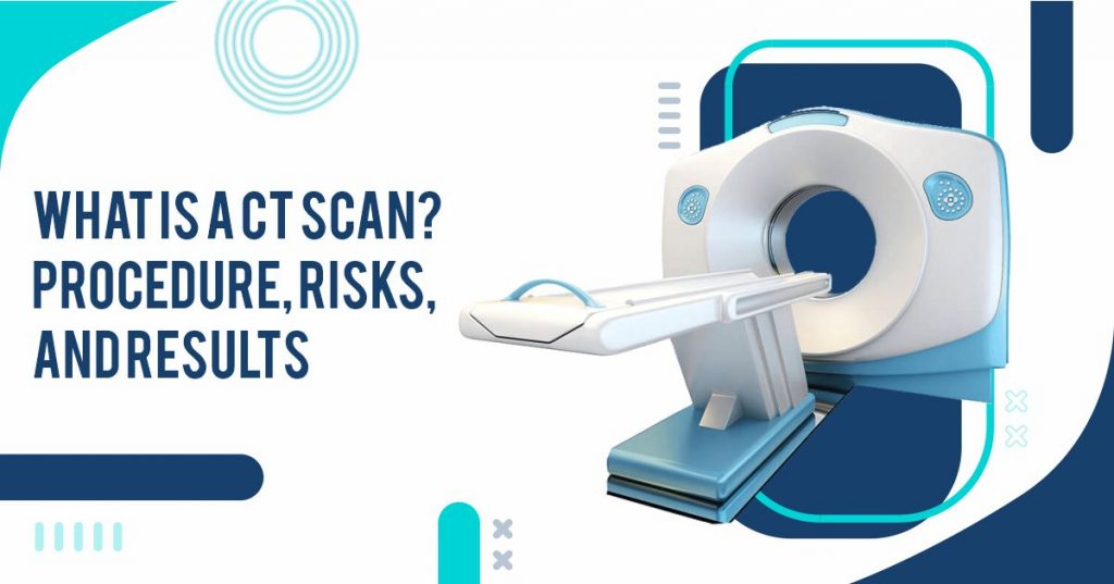
CT machines are typically large, donut-shaped machines with a short tunnel in the center. As you lie on the narrow examination table that slides into the short tunnel during your surgery, your image is taken. In the center of the room, opposite each other in a ring, are an x-ray tube and an electronic x-ray detector. A computer is located in a control room where the images are processed.
The technologist operates a scanner and monitors your exam, watching it directly from your side. A speaker and microphone will allow the technologist to speak to you.
How Do CT Scans Work?
CT scanners do not use conventional x-ray tubes; instead, they are powered by a motorized x-ray source that rotates around a circular opening that is built into a donut-shaped structure called a gantry.
CT scans or CAT scans process involves a patient lying on a bed moving slowly through a gantry while an x-ray tube rotates around the patient, traveling through narrow beams of light. Towards the end of the x-ray, the detectors detect it and transmit it to the computer.
You Can Read Also: What Is Atherosclerosis?
During each rotation of the x-ray source, the CT computer constructs a 2D image slice of the patient utilizing sophisticated mathematical techniques. The thickness of tissues showed in each slice image varies slightly by type of CT machine but usually ranges from 1-10 millimeters. After a complete image slice has been obtained with the help of a scanning machine, the image is stored and the motorized bed is incrementally moved forward into the gantry.
After that, an additional x-ray scan is conducted to produce another slice, which is repeated until the desired number of slices is accumulated. An image obtained by the CT scan whose slice can be displayed either individually or stacked together by the computer as a 3D image of the patient. This 3D image shows the skeleton, organs, and tissues of the patient as well as any abnormalities that may be present.
Among its many advantages, this method allows you to rotate the 3D image in space or view slices sequentially, which makes it easier to locate the exact problem location.
What are the benefits?
- With a CT scan, an experienced radiologist can diagnose many abdominal pain and trauma conditions with a high level of accuracy and enable more effective treatment. It may even eliminate additional, more invasive diagnostic testing.
- The method of a CT examination can be used to decrease the risk of serious complications like those caused by an infected fluid collection or a burst appendix and the subsequent spread of infection when pain is caused by infection and inflammation.
- In addition to being non-invasive and painless, CT scans are accurate.
- A major advantage of CT imaging is that soft tissue, blood vessels, and bone can all be visualized simultaneously.
- With CT scanning, detailed images of bones, lungs, and blood vessels are obtained without the use of conventional x-rays.
- CT scans are simple and fast. In emergencies, they can show internal bleeding and injuries quickly enough to save lives.
- The cost-effectiveness of CT has been demonstrated for a wide range of clinical problems. The sensitivity of CT to patient movement is less than that of MRI.
- In other terms, a CAT scan provides real-time imaging, making it ideal for performing minimally invasive procedures such as needle biopsies and needle aspirations of many parts of the body, including the lungs, the abdomen, the pelvis, etc and the bones.
- CT scanning may allow a direct diagnosis to be made without requiring exploratory surgery or surgical biopsy.
- An X-ray used in a CT scan should not cause any side effects to the patient immediately after the scan. There is no radiation left in the body after the CT scan.
What happens during a CT scan
CT scans are performed lying on your back on a flatbed that passes through a rotating circle as you pass through the scanner. During the scan, you will usually lie on your back in the ring-shaped scanner.
You do not have to worry about feeling claustrophobic since the scanner does not surround your entire body at once. Radiographers will operate scanners in separate rooms. You will have the ability to speak and hear them over an intercom while the scan takes place.
During each scan, you must lie completely still and breathe normally. So that, the scan images will not be blurred. At certain points, you may be asked to take a deep breath, let it out, or hold it. The scan normally takes about 10 to 20 minutes.
What happens afterward
A CT scan is not expected to cause any lasting effects, and most patients can return home shortly afterward. You can drive, eat, and drink as normal.
The contrast used in the test is harmless and passes out of your body through your urine at the end of an hour, so you may be asked to wait in the hospital for up to that long.
You Can Read Also: BAD BREATH: CAUSES, TREATMENTS, AND PREVENTION
You won’t receive the results of your scan instantly. A computer will need to process the information and then examine the picture. A radiologist (a physician who interprets images of the body) will then interpret the results.
An MRI radiologist examines your images and develops a report he or she sends to the referring doctor so that you can discuss the results with them. This usually takes a few days or weeks to complete.
What Is It Used For?
CT scan is useful for obtaining images of:
- soft tissues
- blood vessels
- lungs
- the pelvis
- brain
- abdomen
- bones
The CT scan is often preferred for diagnosing many types of cancer, including liver, lung, and pancreatic cancer. It provides clear images that allow doctors to determine how much tissue is affected by the tumor, its size, and its location.
The results of a CT scan can reveal a tumor in the abdomen and any swelling or inflammation in nearby organs if there is a tumor in the head. A head scan can reveal bleeding, swelling of the arteries, or a tumor on the brain.
A CT scan can provide valuable information on blood flow, kidneys, liver, and spleen lacerations. Also, a CT scan detects abnormalities in tissue, making it useful for planning radiotherapy and biopsies.
A doctor can use it to assess bone disease and density and the condition of a patient’s spine. Additionally, it provides vital information about injuries to the hand, foot, or other skeletal structures of a patient. The surrounding tissue around small bones can even be seen.
What Are the Risks obtained through CT Scan?
It is considered that radiation exposure during a CT scan is low, so if you are pregnant or believe that you may be pregnant, you should notify your doctor immediately.
A patient with a history of hypersensitivity to contrast dye, shellfish, medications, or iodine should tell their physician if they have any of these allergies. Radiation exposure during pregnancy contributes to birth defects.
It is important to notify your physician if you have kidney failure or another kidney disease. Contrast dye can sometimes cause kidney failure, especially when the person is taking Glucophage (a diabetic medication).
A chest CT scan may be more accurate if certain factors interfere with the accuracy of the procedure. Your physician should be consulted about any concerns you have about the procedure.
There are a variety of factors that contribute to these findings, including, but not limited to:
- Surgical clips or pacemakers, for example, maybe, metallic objects inside the chest
- Body piercings on the chest
- A recent barium study in the esophagus showed barium in the organ
What Do CT Scan Results Mean?
If the radiologist doesn’t see tumors, blood clots, fractures, or other abnormalities in the scan images, it may require further tests or treatments. If abnormalities are detected, further tests or treatments may be needed, depending on the type of abnormality.

