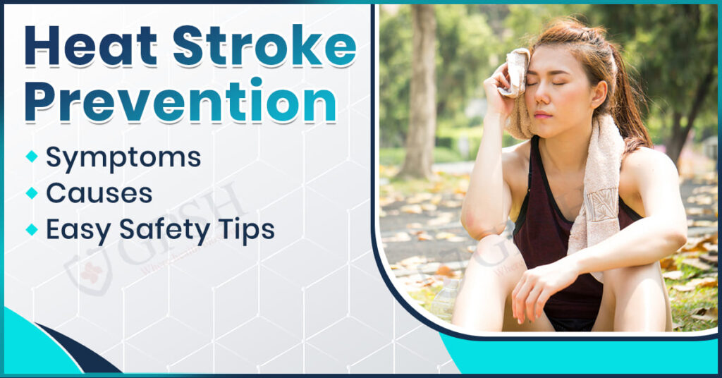
Sarvesh Joshi

Obstructive Sleep Apnea

Obstructive Sleep Apnea (OSA): Causes, Symptoms, Risk and Prevention Options

Introduction

Obstructive Sleep Apnea (OSA) is a prevalent but severe sleep disorder in which the muscles of the throat become too relaxed, causing repeated interruptions in breathing while sleeping. Such pauses during breathing result in fragmented sleep, reduced levels of oxygen, and severe health complications if not treated. In this article, causes of OSA such as anatomical causes, obesity, and habits will be discussed. We will also cover its signs, like boisterous snoring, excessive daytime tiredness, and a lack of focus. Moreover, we will analyze the risk factors that lead to the onset of OSA and its potential effects on overall health. Finally, we will summarize different preventive measures and therapeutic solutions, varying from lifestyle adjustments and medical therapies to innovative treatment modalities.

What is Obstructive Sleep Apnea (OSA)?

Obstructive Sleep Apnea (OSA) is a sleep disorder that involves frequent episodes of partial or total obstruction of the upper airway during sleep, resulting in disrupted breathing, decreased oxygen levels, and disturbed sleep patterns.

Causes of Obstructive Sleep Apnea

Obstructive Sleep Apnea (OSA) refers to states within the body that cause occlusion or narrowing of the airway during the hours of sleep. The major causes include:

  • Excess Weight & Obesity: Fat around the neck and throat could limit airflow.
  • Anatomical Factors: Increased size of tonsils, an enormously big neck, recessed jaw, or narrow airway can also increase risk.
  • Muscle Relaxation: It could lead to the collapse of the airway due to excessive relaxation of muscles in the throat during sleep.
  • Aging: A decline of muscle tension in the throat associated with age can provide the condition of causing airway obstruction.
  • Genetic Predisposition: The occurrence of OSA in families could make a person more susceptible to developing the condition.
  • Nasal Restriction and Blockages: Nasal congestion that occurs repeatedly, septal deviation, or allergies may restrict airflow.
  • Alcohol or Sedatives: These relax throat muscle tissues. Therefore, they are predisposing factors to malocclusion of airways.
  • Smoking: The situation will commit the air passage to an effective narrowing due to both the inflammatory process and the accumulation of fluid within an upper airway.
  • Hormonal Imbalances: OSA can also be induced by conditions such as hypothyroidism and polycystic ovary syndrome (PCOS).

You can read also:- Sialendoscopy: What are Causes, Symptoms, Preventions, and Treatment Options

Symptoms of Obstructive Sleep Apnea

Obstructive Sleep Apnea (OSA) has a way of ruining sleep and health, manifesting through signs and symptoms.

  • some common symptoms:
  • Loud Snoring: Accompanied by gasping or choking noises typically noted by other people.
  • Breathing Pauses: The breathing stops for several seconds to minutes at night, which may be commented on by the partner.
  • Daytime Sleepiness: Unusually, tiredness persists during the daytime hours even when a night’s sleep is adequate.
  • Morning Headaches: These are caused by low oxygen levels during sleep.
  • Trouble Concentrating: Unable to focus, forgetful, and it is struggling to perform day-to-day activities.
  • Mood Changes: Excessively irritable, stressed, anxious, or feeling down.
  • Dry Mouth or Sore Throat: Often occurs because of breathing through the mouth while sleeping.
  • Waking Up Frequently at night: Sometimes with a choking feeling.
  • Nocturia, or Frequent Nighttime Urination: Waking up several times for bathroom use.
  • Long-Term Consequences:
    • High Blood Pressure (Hypertension): Repeated oxygen drops put a strain on the cardiovascular system.
    • Heart Disease & Stroke: Risk is increased because of inadequate oxygenation and blood pressure surges.
    • Type 2 Diabetes: OSA is associated with insulin resistance and metabolic syndrome.
    • Liver Issues: Fatty liver disease and abnormal liver function tests are prevalent.
    • Compromised Immune Function: Repeated inflammation and impaired body repair mechanisms.
    • Greater Risk of Accidents: Because of daytime sleepiness and reduced alertness.
    • Cognitive Decline: Memory impairment and heightened vulnerability to dementia in advanced cases.

Risk Factors for Obstructive Sleep Apnea

Multiple conditions enhance the possibility of getting Obstructive Sleep Apnea (OSA). These include:

  • Obesity: Too much fat in the neck region may clog air passage.
  • Large Neck Circumference: More than 17 inches in men and 16 inches in women poses increased risk.
  • Small Airway: Hereditary or acquired factors such as a small jaw, big tonsils, or large tongue.
  • Nasal Obstruction & Congestion: A deviated septum, constant allergies, or sinuses can narrow airflow.
  • Smoking: Induces inflammation and water retention in the upper airway.
  • Alcohol & Sedative Use: Suppresses throat muscle tone, elevating airway collapse risk.
  • Poor Sleep Positioning: Sleeping on the back may exacerbate airway obstruction.
  • Aging: Muscle tone diminishes with age, raising airway collapse risk.
  • Hormonal Imbalances: Disorders such as hypothyroidism and PCOS are associated with OSA.
  • Diabetes: Individuals with type 2 diabetes are at greater risk for sleep apnea.
  • Heart Disease & Hypertension: Cardiovascular conditions are a risk factor for OSA, as well as one of the outcomes.
  • Family History: Those who have close family members who have OSA are at increased risk.
  • Gender: Males have a greater likelihood of acquiring OSA, although postmenopausal females have an increasing risk.

You can read also:- Endocarditis: Causes, Symptoms, Risk Factors, and Treatment

Prevention Options for Obstructive Sleep Apnea

Elimination of risk factors for developing OSA and severity is considered preventive management. Important preventive strategies include:

  • Keep Weight Under Control: Losing weight can reduce fat deposits in the neck, which helps in moving air. Exercise maintains body fitness and also tones the muscles of the airway.
  • Sleep Positioning: Side sleeping is better than back sleeping because it prevents airway collapse. A positional therapy device or wedge pillow may help to keep one in a side-sleeping position.
  • Avoid Alcohol & Sedatives: Alcohol and sedatives relax throat muscles, which increases the chances of airway obstruction. Therefore, these should be avoided or minimized before sleep to improve one’s breathing.
  • Quit Smoking: Smoking incites inflammation and water retention of the upper airway. Quitting might decrease airway swelling and allow for better oxygen flow.
  • Treat Nasal Congestion: Treatment of allergies, a sinus infection, or a deviated septum opens the nasal passages. Decongestants or nasal sprays, antihistamines, or humidifiers will aid in allowing free airflow.
  • Encouraging good sleeping habits: Get 7-9 hours of quality sleep each night; maintaining these time schedules will synchronize your natural sleep-wake cycle.
  • Strengthening the throat muscles: Activities such as singing or playing wind instruments can help improve the tone of the airway muscle, myofunctional therapy.
  • Routine Health Check-Ups: Look for conditions such as high blood pressure, diabetes, or hormonal imbalance that could give way to OSA. Also, if loud snoring and daytime fatigue are observed, seek early consultation with a doctor.

Conclusion

Obstructive Sleep Apnea (OSA) is a serious disorder of sleep that can heavily affect general health if not treated. It is due to states of obesity, anatomical deformity, and lifestyle, resulting in symptoms of snoring loudly, daytime sleepiness, and susceptibility to cardiovascular illness. Early diagnosis and treatment are essential, and prevention through control of weight, sleeping position, and lifestyle alteration. OSA is usually managed in Pulmonology, Sleep Medicine, and ENT (Ear, Nose, and Throat) departments by specialty doctors providing innovative treatment. In order to experience professional treatment, Shekhawati Hospital provides state-of-the-art diagnosis centers and best therapies to guide the patients towards quality sleep as well as better overall health.

Obstructive Sleep Apnea (OSA): Causes, Symptoms, Risk and Prevention Options Read More »

Sialendoscopy

Sialendoscopy: What are Causes, Symptoms, Preventions, and Treatment Options

Introduction

Sialendoscopy is a minimally invasive procedure indicated for the diagnosis and treatment of salivary gland disorders which are commonly caused by obstructions like salivary stones, strictures, or infections. These conditions contribute to swellings, pain, and difficulties in saliva flow which commonly lead to debilitations of oral health and comfort.

In this article, we will discuss the different kinds of causes through which salivary gland obstruction occurs, its symptoms which reflect a medical problem, types of preventive measures available to avoid further glandular difficulties, and modes of treatment- one such an example being the sialendoscopy process, which seems to have impressive results. The factors listed out help in providing early diagnosis, and subsequently with management, make them suitable for better long-term oral health maintenance.

What is Sialendoscopy?

Sialendoscopy is a minimally invasive diagnostic and therapeutic procedure for examination and treatment of salivary duct disorders, including sialolithiasis, ductal strictures, and chronic infections. It involves the insertion of a miniature endoscope (0.8–1.6 mm in diameter) into the salivary ducts.

It allows for direct visualization and intervention without external incisions, maintaining gland function while enabling stone removal, ductal dilation, and lavage therapy. Biologically, sialendoscopy promotes the salivary gland by reinstating normal salivation flow and decreasing inflammation as well as the recurrence of infections.

You can read also:- Endocarditis: Causes, Symptoms, Risk Factors, and Treatment

Causes of salivary gland disorders

Here are the very few causes of Salivary Gland Disorders Leading to Sialendoscopy.

  • Sialolithiasis (Salivary Stones): Calcium-based deposits obstruct salivary ducts, thus causing swelling and pain.
  • Ductal Strictures: These are narrow bands of salivary ducts caused by scar tissue, inflammation, or recurrent infections.
  • Chronic Sialadenitis: Recurrent infections from bacteria and viruses cause constant gland swelling and dysfunction.
  • Autoimmune Disorders: Such conditions like Sjӧgren’s syndrome lead to decreased saliva secretion and duct blockages.
  • Mucous Plugs: The plugs of thickened saliva or mucus block ducts, giving a sense of pain and inflammation.
  • Trauma or Surgery: Ducts become scarred and obstructed by previous surgical interventions, injury, or radiotherapy.
  • Dehydration and Impaired Saliva Flow: Lack of saliva raises the risk for stone formation as well as for infection.
  • Drugs: Inhibitors to saliva production: antihistamines, diuretics, anticholinergics.
  • Tumors or Cysts. Benign and malignant tumors might compress the ducts of their glands.

Symptoms of Salivary Gland Disorders

Here are some of the symptoms of Salivary Gland Disorder.

  • Swelling of the Salivary Glands – Repeated or persistent swelling, often near the jaw, cheeks, or under the tongue.
  • Pain or Tenderness – Pain, particularly when eating or drinking, as a result of duct obstruction.
  • Dry Mouth (Xerostomia) – Decreased saliva production, causing difficulty in chewing, swallowing, or speaking.
  • Obstruction of Saliva Flow – Feeling of blocked or decreased saliva production.
  • Recurrent Infections – Infection of the ducts repeatedly by bacteria or viruses, giving rise to redness, warmth, and discharge of pus.
  • Bad Taste or Foul Smelling Discharge – Pus or bacterial collections causing an objectionable taste or odor in the mouth.
  • Hard Lumps or Stones in the Gland Area – Palpable stones present within the salivary ducts.
  • Fever and General Malaise – Systemic signs of infection can occur, especially fever and generalized malaise.
  • facial or jaw pain. It may radiate to other surrounding areas and may worsen with meals.

How is Sialendoscopy Performed?

Steps of Performing Sialendoscopy

  • Preparation of the Patient

    • Local or general anesthesia is provided depending on the severity of the procedure.
    • The salivary duct opening is softly dilated for scope placement.
  • Placement of Sialendoscope

    • A micro-endoscope with a diameter of 0.8–1.6 mm is inserted slowly into the salivary duct for viewing purposes.
  • Duct Examination

    • Camera and fiber-optic light give a magnified view of the interior of the duct. Any blockage present, such as stones, strictures, or infections, is detected.
  • Procedure for Treatment

    • Salivary Stone Removal – The stones are removed using micro forceps, baskets, or laser fragmentation.Ductal Dilation – Tight strictures or narrowing is dilated by small balloon catheters.
    • Lavage Therapy – Saline or anti-inflammatory solutions flush out debris and infection in the duct.
  • Final Inspection and Scope Removal

    • The duct is checked for clearance and the scope is removed with care.
  • Post-Procedure Care

    • Patients are monitored for swelling or discomfort.
    • Hydration and gland massage are promoted to stimulate salivation.
    • In case of necessary, antibiotics or pain relievers may be given.

You can read also:- Neck Pain in Children: Causes, Symptoms, and Treatment

Treatment Options for Salivary Gland Disorders

Disorders of the Salivary glands are caused due to infections, blockages, autoimmune diseases, or tumors. This condition depends upon the cause as well as on the severity; hence, some of the chief treatment options follow:

  • Medications & Conservative management
    • Antibiotics: When there is an infection caused by bacterial pathogens (for example sialadenitis).
    • Anti-inflammatory & Analgesics- NSAIDs include ibuprofen which helps minimize pain and edema.
    • Liquids and Sialogogues- More intake of fluid and sucking some sour candies sialogogues help stimulate salivary gland flow.
    • Massage & Warm Compress: Facilitate dislodgement and allow gland drainage.

  • Treatments of Obstruction
    • Manual massage and hydration: Stones can be expectorated through the mouth
    • Sialendoscopy: Minimally invasive removal of salivary stones
    • Surgical removal: For large and embedded stones.

  • Treatment of Autoimmune Conditions like Sjögren’s Syndrome
    • Artificial saliva & eye drops: They facilitate moisturization.
    • Medications: Pilocarpine or cevimeline increases the amount of saliva.
    • Immunosuppressants: Steroids or biologics in severe cases

  • Treatment of Tumors Benign or Malignant
    • Surgical removal: First-line treatment for a tumor (such as pleomorphic adenoma)
    • Radiation therapy: Malignant tumors, or if surgical intervention is not possible
    • Chemotherapy: Not very common; it is utilized in aggressive and metastatic conditions

  • Treatment of Viral Infections such as Mumps
    • Supportive care: Rest, hydration, and pain control by NSAIDs or acetaminophen
    • Isolation & prevention: Mumps vaccination, MM End.

Preventing Salivary Gland Issues

Here are some precautions you need to take so you do not get Salivary Gland Issues.

  • Keep Your Mouth Hygienic: Brushing at least twice daily, flossing, and using an antibacterial mouthwash help prevent bacteria in the mouth. Poor oral hygiene can create a buildup of bacteria, which can travel down to the salivary glands and cause infection like sialadenitis which is inflammation of the salivary gland.

  • Stay Hydrated: Drinking enough water throughout the day keeps the salivary glands active and prevents dehydration. A dry mouth increases the risk of bacterial infections and salivary gland blockages, which may lead to conditions such as xerostomia or dry mouth syndrome. Limit your intake of caffeine and alcohol, as these are dehydrating agents that decrease saliva production.

  • Stimulate Saliva Flow: Chew sugar-free gum or eat foods high in fiber (such as apples, carrots, and celery) to naturally stimulate saliva. Saliva serves to wash out bacteria and food particles and also prevents the development of salivary stones.

  • Avoid Smoking and excessive Alcohol. Smoking and alcohol irritate the salivary glands, increasing the chance of infection and blockage. These habits may also contribute to decreased saliva production, which can cause dry mouth and bacteria growth.

  • Prevent Salivary Stones: Salivary stones, also known as sialolithiasis, are formed when saliva becomes concentrated, resulting in crystallized deposits in the glands. A balanced diet rich in fluids and low in excessive calcium intake helps prevent stone formation. Gently massaging the salivary glands can promote saliva flow and reduce blockages.

  • Manage Underlying Conditions: Several medical conditions lead to decreased production of saliva by the salivary glands; these include diabetes, Sjögren’s syndrome, and autoimmune diseases. Management of the underlying conditions either through medication or lifestyle adjustments safeguards the salivary glands against dysfunction.

  • Protect Against Viral Infections: Viral infections, including mumps (which are caused by the mumps virus), influenza, and cytomegalovirus, directly affect the salivary glands. Getting vaccinated with the MMR vaccine (measles, mumps, and rubella) and maintaining good hygiene (frequent handwashing and avoiding contact with infected individuals) helps prevent these infections.

Conclusion

Salivary gland disorders impair oral and general health, often resulting from infections, blockage, autoimmune conditions, or sometimes due to tumors. Of the most advanced and minimally invasive procedures to treat salivary gland disease, especially salivary duct stones, is sialendoscopy.

This procedure lets a doctor come to your rescue with precise diagnosis and treatment that brings less discomfort and quicker recovery than surgery. Salivary gland disorders and their treatments are undertaken in the departments of Otolaryngology (ENT) and Oral & Maxillofacial Surgery, where specialized care is provided. Shekhawati Hospital has a reputation for diagnosing and treating salivary gland conditions with advanced procedures such as sialendoscopy to ensure effective and patient-friendly treatment.

Sialendoscopy: What are Causes, Symptoms, Preventions, and Treatment Options Read More »

Endocarditis

Endocarditis: Causes, Symptoms, Risk Factors, and Treatment

Introduction

Endocarditis is an infection of the inner lining of the heart chambers and valves. It is caused by bacteria or other pathogens entering the bloodstream and attaching to heart tissues. It can lead to severe complications if not treated promptly, including heart failure and the spread of infection to other organs. In this article, we will explain what causes endocarditis, symptoms to watch out for, the main risk factors for the disease, and how to treat the condition. In addition, we shall discuss diagnostic procedures used to determine endocarditis and prevention procedures to prevent the infection.

What is endocarditis?

Endocarditis is an infection of the endocardium, the inner lining of the heart chambers and valves, often caused by microorganisms such as bacteria, fungi, or viruses. The infection occurs when these pathogens enter the bloodstream, either through bacteremia or fungemia, and adhere to damaged or abnormal heart tissue. Infection leads to inflammation, causing damage to the heart valves and disrupting normal cardiac function. Severe consequences of endocarditis include heart valve destruction, embolism, and septicemia. Thus the early detection and treatment of endocarditis are of essence for survival.

You can read also:- Neck Pain in Children: Causes, Symptoms, and Treatment

Who does endocarditis affect?

It may only affect those that have heart anomalies, but actually, endocarditis can result in any of the population without any distinction whatsoever. The categories of individuals include:

  • Heart Abnormalities in People: All those who happen to have intrinsic heart malformation or broken down valves in the heart due to their rheumatic conditions are prone.
  • Those with Artificial Valve in Heart People: All such people that undergone heart valve substitution surgery or patients with artificial hearts may readily be infected with the condition.
  • Individuals with a Previous History of Endocarditis: Those who have had endocarditis before are more likely to have it in the future.
  • Immunocompromised People: People whose immune system is weakened, including those suffering from HIV/AIDS, cancer, and individuals on immunosuppressive medications, are also at risk.
  • Intravenous Drug Abusers: People using intravenous drugs with non-sterile needles are at very high risk as pathogens may directly enter their bloodstream.
  • People with specific medical conditions are more susceptible to developing endocarditis; these include diabetes, kidney disease, or any illness that demoralizes a patient’s immune system.
  • Old age: Generally, there can be age-related changes or certain diseases associated with older age.
  • Dental or surgical procedures: Patients undergoing invasive procedures, especially those concerning the heart, teeth, or urinary tract, are at increased risk if precautions are not properly taken.

How common is endocarditis?

Endocarditis is relatively uncommon, but its occurrence has been steadily increasing in recent years due to the increasing numbers of people afflicted with heart disorders and prosthetic heart valves. In the United States, the estimated annual incidence of infective endocarditis is placed at 3 to 10 cases per 100,000 people. Higher incidences often occur in a specific population that includes:

  • Older adults: With the aging population, the incidence of endocarditis in people over 65 years has been increasing.
  • Intravenous drug users: Due to increased drug usage and risks, such as needle sharing with no sterilization.
  • Individuals with prosthetic heart valves or congenital heart defects: In this category, the risk is higher because of structural abnormalities in the heart.

What are the causes of endocarditis?

It is primarily caused by infection from bacteremia or fungemia, which is due to bacteria, fungi, or other pathogens that attach themselves to the inner lining of the heart called the endocardium. The most common causes of endocarditis include:

  • Bacterial Infections
    • Streptococcus species: The most common causative agent of endocarditis is the bacteria, especially Streptococcus viridans, which comes from dental procedures or oral infections.
    • Staphylococcus aureus: This bacterium, which includes methicillin-resistant Staphylococcus aureus (MRSA), is one of the most common causes, frequently related to the use of intravenous drugs or healthcare-acquired infections.
    • Enterococci: The bacteria inhabit the GI and urinary tracts but can cause endocarditis in people with heart disease or who are receiving invasive medical procedures.
    • Other bacteria, like Streptococcus bovis (associated with colon cancer), Haemophilus, Actinobacillus, Cardiobacterium, and Eikenella, which are known together as the HACEK group, can cause endocarditis, but much less frequently.
  • Fungal Endocarditis
    • Fungal endocarditis is much less common, but it may occur, especially in immunocompromised patients or in individuals who have long-term intravenous catheters. Candida species are the most common fungi causing endocarditis.
  • Intravenous Drug Use
    • The use of non-sterile needles to inject drugs may directly introduce bacteria or fungi into the bloodstream, thus raising the risk of endocarditis. Staphylococcus aureus, including MRSA, is often implicated in endocarditis among intravenous drug users.
  • Healthcare-Associated Infections
    • Intra-vascular procedures such as heart valve surgery, catheter insertion, and dialysis, may introduce pathogens into the blood, especially in a person with a previous history of heart conditions or a compromised immune system.
  • Dental and Oral Infections
    • Poor oral hygiene, dental procedures, or gum disease (periodontal disease) can allow bacteria, including Streptococcus viridans, to enter the bloodstream and then infect the heart valves.
  • Underlying heart conditions
    • Structural heart abnormalities or valve diseases, such as congenital heart problems, rheumatic heart disease, or damaged valves, predispose the heart to easily attach pathogens, thereby exposing it to infection.

You can read also:- What is Tinnitus? Causes, Symptoms, Diagnosis and Treatment

Symptoms of endocarditis

The symptoms of endocarditis are quite variable based on the intensity of the infection, which heart valve is affected, and the health status of the patient. Some common symptoms include:

  • Recurring fever and chills: Most common signs of infection.
  • Fatigue and weakness: General tiredness and weakness due to the body fighting off the infection.
  • Heart murmur: A new or changed heart murmur can be observed by a doctor.
  • Shortness of Breath: Inability to breathe due to inefficient pumping of the heart.
  • Swelling: Swelling in the legs, feet, or abdomen caused by fluid retention.
  • Chest Pain: Chest pain or discomfort, which could be a sign of heart damage.
  • Coughing (sometimes with blood): Lung involvement may cause coughing that potentially contains blood.
  • Unexplained Weight Loss: Infection that is persistent and results in weight loss for an unknown reason.
  • Germanic Symptoms – Petechia, Osler’s node, Janeway’s lesion, and splinter hemorrhages on the cutaneous level.
  • Persistent or Severe Headaches: Headaches are caused by an infection that has impacted the brain.
  • Blood in the urine: blood in the urine due to kidney involvement.
  • Joint and Muscle Pain: Inflammation causes pain in joints and muscles.

Risk factors for endocarditis

Endocarditis is an infection of the heart’s inner lining, often including the heart valves. The risk factors for endocarditis are:

  • Heart Valve Abnormalities: Congenital defects, rheumatic heart disease, or damaged heart valves increase susceptibility.
  • Prosthetic Heart Valves: Individuals with artificial heart valves are at higher risk of infection.
  • Previous Endocarditis: A history of endocarditis increases the likelihood of recurrence.
  • Intravenous drug use: Using a non-sterile needle may increase the chances of bacterial inoculation.
  • Immunocompromised states: States that cause weakening of the immune system such as HIV/AIDS, cancer, or immunosuppressive therapy.
  • The elderly are more susceptible because of the aging changes in the heart and other associated conditions.
  • Invasive medical procedures: Such infections arise after open-heart surgery, dental surgery, or a catheter insertion in almost every body organ.
  • Even chronic illness, say, diabetes, kidney disease, or any other systemic disease increases vulnerability.
  • Poor Oral Hygiene or Gum Infection: Dental infection or dental procedures that may invade the oral cavity may expose the heart to bacteria.
  • Urinary or Gastrointestinal System Infections: Infections in these systems may spread bacteria through the bloodstream to the heart.
  • Close contact with an infected person: Close contact with a diseased person may increase the risk, especially if there is a compromised immune system.

Management and Treatment

The management and treatment of endocarditis are to eliminate the infection, prevent complications, and manage underlying diseases. The approach for treatment depends on the causative organism of the illness and the severity of the disease. This is an extensive outline:

  • Antibiotics:
    • Empiric therapy: broad-spectrum intravenous antibiotics (such as penicillin, and vancomycin) until the organism cultures are available
    • Targeted therapy: antibiotics are adjusted according to the organism; e.g., a patient infected with Staphylococcus aureus would be treated with nafcillin or vancomycin.
    • Duration: Generally 4-6 weeks of IV antibiotics.
  • Surgical Intervention:
    • Severe damage or infection with valve repair or replacement.
    • Drainage of abscesses or removal of infected prosthetic devices, if required.
  • Supportive Care
    • Hospitalization with IV antibiotics and monitoring
    • Monitoring heart function with echocardiograms
    • Management of pain for the associated discomfort with the infection
  • Prevention
    • Administration of prophylactic antibiotics before dental or invasive procedures for at-risk patients
    • Good oral hygiene to prevent bacterial entry
    • Good management of predisposing conditions such as diabetes, heart disease, or intravenous drug use.
  • Monitoring for Complications
    • Heart Failure: Monitor for signs of heart failure due to valve damage.
    • Stroke: Monitor neurological symptoms indicating embolic events.
    • Kidney Failure: Regularly assess renal function.
    • Sepsis: Be on the lookout for systemic infection signs.
  • Long-term Follow-up:
    • Regular Monitoring: Echocardiograms are to be followed up periodically for the resolution of infection.
    • Complete Antibiotic Course: To avoid relapse, complete the prescribed antibiotics.

Conclusion

Endocarditis refers to a life-threatening infection of the lining of the heart also called endocardium, whose immediate detection and treatment are required to forestall grave sequelae like heart failure, stroke, or visceral organ damage. Usually ended through antimicrobial therapy and surgical intervention with sustained supportive care. Besides, preventative steps comprising antibiotics and an ever-favorable oral condition are significant for high-risk individuals. Infection warranting the expertise of an infectious disease specialist, endocarditis falls within the realm of Cardiac Sciences. To deliver holistic care and the very finest treatment options possible, if one has endocarditis, the Shekhawati Hospital in Jaipur happens to be one of the best. With modern medical facilities coupled with a committed, brilliant, and highly efficient cardiology team, the center offers an opportunity to experience the best outcome for its patients.


Endocarditis: Causes, Symptoms, Risk Factors, and Treatment Read More »

Neck Pain

Neck Pain in Children: Causes, Symptoms, and Treatment

Introduction

Once considered a rare complaint, neck pain in children has become a concern in today’s world because of changing lifestyle factors, increased screen time, and demanding school routines. Neck pain may seem to be a minor issue, but it may indicate some issues that need attention to prevent long-term complications.

This article focuses on neck pain in children, from poor posture and sports injury to medical conditions like infections and congenital anomalies. We’ll cover the symptoms that parents should be aware of and how to diagnose the problems, as well as treatment options ranging from lifestyle changes and physical therapy to medical interventions. As the early stage of the children’s physical condition is very vulnerable, one should not ignore neck pain at the earliest so that this should not hamper their overall development or health.

What is neck pain?

Neck pain is the discomfort or feeling localized to the region of the cervical spine, involving the muscles, nerves, intervertebral discs, ligaments, and vertebrae. Biologically, it is usually related to mechanical stress, inflammation, nerve compression, or degenerative changes in the cervical structures.

You can read also:- Cervical Spine: Anatomy, Diseases, Disorders and Treatments

Why Does Neck Pain Occur in Children?

There are several causes for neck pain in children, including benign to some serious causes. It is generally not a very rare condition but results from some lifestyle habits injuries or any other underlying disease.

Common causes of neck pain in children are as follows

  • Muscle strain or overuse:
    • Causes include poor postures, for instance, slouching over, and hunched over electronic devices.
    • Overuse of a smartphone, a tablet, or a computer may give rise to the condition called “text neck”.
    • Overuse during certain sports or exercises.
  • Injury or Trauma:
    • Neck strain or whiplash may result from falls, accidents, or sports injuries.
    • jerking movements during physical activity
  • Infections:
    • Throat infections such as tonsillitis or pharyngitis cause referred pain in the neck.
    • Lymphadenitis: Swelling of the lymph nodes in the neck caused by an infection.
    • Meningitis: A serious illness that can produce neck stiffness, pain, along with fever.
  • Torticollis (Wry Neck):
    • A condition where the neck muscles contract, causing the head to tilt to one side.
    • May be due to muscle strain, birth injuries, or idiopathic reasons.
  • Congenital Conditions:
    • Structural deformities of the cervical spine may result in chronic neck pain.
    • Include Klippel-Feil syndrome, fusion of cervical vertebrae
  • Psychosocial Factors:
    • Anxiety or tension may cause the contraction of neck muscles.
  • Poor sleep habits
    • Slept awkwardly or improper pillows may stretch neck muscles
  • Heavy school bags
    • Heavy load of schoolbag may cause overstretching of neck and shoulder muscles
  • Juvenile Rheumatoid Arthritis:
    • Inflammatory diseases such as arthritis can cause neck pain, stiffness, and swelling.
  • Spinal Conditions:
    • Diseases like scoliosis or herniated cervical discs are relatively rare but may cause pain.

Symptoms of Neck Pain in Children

Neck pain symptoms in children differ from the actual cause. These are some common symptoms that point to neck pain:

General Symptoms:

  • Pain or Discomfort: Pain located in the neck, which may be dull, sharp, or throbbing in nature. Such pain often gets worse with movement or other related activities.
  • Stiffness: One may not easily turn or move the neck, with a sense of tightness or a loss of range of motion.
  • Tenderness: Pain upon the application of pressure to the neck.
  • Muscle Spasms: Neck muscles involuntarily tighten or cramp.
  • Headache: Pain originating from the neck that can extend into the head, most often a tension-type headache.

Causes and Associated Symptoms:

  • Infectious Causes: (for example, pharyngitis, lymphadenitis, meningitis): Swollen lymph nodes in the neck. Fever, chills, or signs of systemic illness. A stiff neck, especially with meningitis, can also present with headache, nausea, or vomiting.
  • Trauma or Injury: Pain that develops immediately in the neck following a fall or accident. Swelling, bruising, or tenderness in the neck region.
  • Torticollis or Wry Neck: The inability to straighten the neck due to tilting to one side. Poor alignment of the neck.
  • “Text Neck” or Poor Posture: Pain or discomfort following prolonged use of devices. Shoulders are rolled forward or sloping posture.
  • Neurological Symptoms: (less frequent and associated with nerve compression or serious conditions): Paresthesias, weakness, or numbness in the arms or hands; loss of coordination or balance.

You can read also:- Carpal Tunnel Syndrome: Symptoms, Causes, Risk Factors & Treatment

When to See a Doctor?

Seek medical care if the neck pain:

  • Continues for several days or worsens.
  • Accompanies a fever, headache, or stiffness.
  • Occurs following a trauma or injury.
  • Follows neurological signs, such as numbness, tingling, or weakness in the arms.
  • Impairs mobility in the neck considerably.

Home Remedies for Neck Pain in Children

Neck pain in children can often be managed with simple and safe home remedies. These remedies help relieve pain, reduce inflammation, and promote healing without the need for medication in most cases.

  • Rest and Support: Encourage your child to rest and avoid activities that strain the neck, like rough play or prolonged screen time. Use a supportive pillow to keep the neck in a neutral position during sleep.
  • Warm or Cold Compress
    • Cold Compress (First 24–48 Hours): Use an ice pack or frozen peas, cover this with a towel, and place it on the affected area for 15–20 minutes. This will help reduce swelling and numb the area.
    • Warm Compress (After 48 Hours): Use a warm towel, heating pad, or hot water bottle on the neck to relax the muscles and increase blood flow. Repeat this procedure 2–3 times a day for 15–20 minutes.
  • Gentle Massage: Lightly massage the neck muscles using gentle pressure and warm coconut, olive, or lavender oil. This helps to relax tensed muscles and enhance circulation.
  • Stretching and Relaxation Exercises: Once the pain has subsided, teach simple neck stretches: Tilt the head gently from side to side. Slowly rotate the neck in circles. All movements should be slow comfortable and not painful to avoid overstretching.
  • Instill Good Posture: When your child studies, plays, or is on gadgets, teach them to sit straight. The height of the study table and screen may be adjusted accordingly to avoid slouching.
  • Urge Break: Let your child know he needs to take short breaks after 30-40 minutes while engaged in reading, studying, or using gadgets. Get him moving and stretching.
  • Use Epsom Salt Bath: Soak your child in a warm bath containing 1–2 cups of Epsom salt for 15–20 minutes. Magnesium from Epsom salt can help relax the muscles and decrease soreness.

Medical Treatment Options for Neck Pain in Children

Persistent neck pain is rare in children, and when encountered, needs careful evaluation to pinpoint the cause. Appropriate treatment depends on the diagnosis. Here is a broad view of the general treatment the patient may require.

Initial Evaluation

  • History and Evaluation: Evaluate the duration, severity, and any accompanying symptoms (such as fever, trauma, weakness, or neurological symptoms).
  • Diagnostic tests may include X-rays, MRI, CT scans, or blood tests based on the suspected infection, injury, and other possible diagnoses.

Non-Surgical Treatments

  • Pain Medications: Acetaminophen (Paracetamol) or Ibuprofen for lesser and moderate pain. Muscle relaxants can be prescribed in case of spasms.
  • Anti-Inflammatory Agents: NSAIDs such as Naproxen or Diclofenac
  • Antibiotics: In case of suspicion or confirmed infection like meningitis, abscess
  • Corticosteroids: In autoimmune cases of Juvenile arthritis or intense inflammation.
  • Other Medications: If the cause is neural then gabapentin may be prescribed.
  • Physical Therapy: Exercises and stretches to improve neck strength and mobility. Heat or cold therapy to reduce pain and inflammation.
  • Lifestyle Modifications: Changing the posture of the child during activities like studying or using devices. Providing ergonomic furniture and a proper pillow for sleeping.
  • Behavioral Therapy: Counseling or relaxation techniques may help in stress-related neck pain.

Surgical Treatments

Surgery is very rarely required in children with neck pain, except in the following conditions:

  • Infections (for example, abscess): Incision and drainage if there is an abscess.
  • Trauma or Fracture: Stabilization surgery, such as spinal fusion, if the cervical spine is unstable.
  • Tumors or Congenital Anomalies: Resection of tumors or structural anomalies.
  • Herniated disc: Rare in children. Microdiscectomy or decompression surgery would be considered in very severe cases.

Conclusion

Children experience neck pain, though it is rare compared to other issues. Neck pain usually has causes such as strain, injury, infection, or an underlying health condition. When properly investigated and treated, patients often find relief from their pain, which helps avert complications. A variety of drug treatments, along with physiotherapy and lifestyle changes, typically alleviate symptoms and usually do not require surgery, except in a few more severe cases. Most children with neck pain fall under the care of pediatric orthopedics or pediatric neurology, depending on the cause. Shekhawati Hospital is one of the best medical facilities for kids suffering from chronic neck pain, including muscle spasms and other related issues. The hospital has an expert team dedicated to providing advanced care and personalized treatment plans to thoroughly address neck pain and related conditions.


Neck Pain in Children: Causes, Symptoms, and Treatment Read More »

Cervical Spine

Cervical Spine: Anatomy, Diseases, Disorders and Treatments

Introduction

This spinal part is composed of the cervical spine, which is located in the neck region; it contains seven vertebrae that support the head and facilitate movement while protecting the spinal cord. The structure is relatively complex and includes bones, discs, muscles, and ligaments that all work to permit flexibility and stability. This vulnerable system however may suffer a lot in different diseases and disorders; therefore, pain, stiffness, and sometimes neurological complaints, like cervical spondylosis, can cause a serious herniation of discs or even result from spinal stenosis.

The paper outlines the anatomy of the cervical spine, common diseases and disorders that affect the area, methods of diagnosis, possible treatment options, as well as preventive measures. To complete this work, further research into surgical roles in treating these diseases as well as new therapeutic possibilities that would emerge.

What is the cervical spine?

The cervical spine is the uppermost section of the spinal column, consisting of seven vertebrae (C1 to C7) located in the neck. It supports the head, allows for neck movement, and safeguards the spinal cord. This part of the spine is essential for maintaining posture, enabling rotation, flexion, and extension of the neck, and supporting both sensory and motor functions.

You can read also:- Migraine: What It Is, Types, Causes, Symptoms & Treatments

What does the cervical spine do?

The cervical spine performs several significant roles including;

  • Support the Head: It upholds the head and gives mechanical support to the structure.
  • Facilitates movement: It supports the neck, which undergoes several movements such as rotations, flexion in which it bends forward, extension in which it bends backward, and lateral bending, a side-to-side motion.
  • Protect the spinal cord: It protects the nervous system of the spinal cord from damage.
  • Enables Nerve Function: The cervical spine contains nerve pathways that control the movement and sensation of the head, neck, arms, and hands.
  • It helps maintain the proper alignment of the head and neck with the rest of the body.

What diseases and disorders affect your cervical spine?

Several disorders and diseases can affect the cervical spine, leading to pain, stiffness, and neurological issues. Some of them are listed below:

  • Cervical Spondylosis: Age-related wear and tear of the cervical spine’s discs and joints, leading to neck pain and stiffness.
  • Herniated disc: When one of the cervical spine’s discs ruptures or bulges, thus pressing on the nerves that are nearby and therefore causing pain, numbness, or weakness.
  • Cervical Stenosis: Narrowing of the spinal canal in the neck, which compresses the spinal cord and nerves resulting in pain, numbness, or weakness in the arms or legs.
  • Cervical Radiculopathy: Compression or irritation of a nerve root in the cervical spine that causes pain, numbness, or weakness radiating into the shoulders, arms, or hands.
  • Whiplash: This is one of the common soft tissue injuries brought about by a sudden jerking motion in the neck due to car accidents, and so on, bringing about pain, stiffness, and headache.
  • Osteoarthritis: Degeneration of the cartilage in the cervical joint leading to discomfort, swelling, and loss of range of motion.
  • Scoliosis is a curvature of the spine that affects the cervical portion, causing discomfort and misalignment.
  • Infections and Tumors: Other, much rare conditions like spinal infection or tumor may cause it to the cervical spine resulting in severe pain and neurologic symptoms.

What are the other muscles and soft tissues in the neck?

The neck comprises various vital muscles and soft tissues that work together to stabilize the cervical spine and provide movement. Some of the key muscles and soft tissues found in the neck are:

  • Sternocleidomastoid (SCM): This prominent muscle originates from the sternum and clavicle and terminates at the mastoid process behind the ear. It helps rotate and tilt the head and facilitates neck flexion.
  • Trapezius: A large muscle that goes from the base of the skull down the back and shoulders. It helps with neck extension, rotation, and stabilization of the scapula.
  • Levator Scapulae: Found at the back and side of the neck, it helps lift the scapula (shoulder blade) and assists in neck rotation and side bending.
  • Scalene Muscles: Three (anterior, middle, posterior) muscles along the lateral sides of the neck provide assistance in breathing and provide flexion to the neck and lateral bending.
  • Deep Neck Flexors: These include the longus colli and longus capitis, muscles deeper within the neck that assist in stabilizing the neck during its primary movement of flexion.
  • Suboccipital Muscles: The small muscles in the posterior base of the skull help to control movements of the head and neck.
  • Platysma: A thin, superficial muscle that runs from the upper chest to the jaw and is used in facial expressions and tension in the neck.
  • Cervical Ligaments: The neck is stabilized through various ligaments, with the anterior and posterior longitudinal ligaments, ligamentum flavum, as well as the nuchal ligament, serving to stabilize and limit too much movement.
  • Tendons and Fascia: These structures support the muscles and help in movement as well as stability by giving attachment of muscles to the bones and an elastic framework for the neck.

You can read also:- ACL Injuries: Causes, Symptoms, Diagnosis & Treatment

How cervical spine diseases can be diagnosed?

The diagnosis of cervical spine disorders is a combination of clinical evaluation and an imaging test. The approach involves:

  • Medical History and Physical Exam: The practitioner examines symptoms, and conducts an examination of the range of motion, strength, reflexes, and signs suggestive of nerve compression or lack of proper alignment.
  • Imaging Tests:
    • Xrays: to identify fractures arthritis, or any alignment issue.
    • MRI-soft tissues such as disks, nerves, and cords are seen in detail.
    • CT Scan: Detailed images of the bones in case of fractures or osteoarthritis.
    • Myelogram: A contrast dye study in case of spinal cord compression or disc problems.
  • Electromyography (EMG) and Nerve Conduction Studies: Determines the function of nerves and helps in diagnosing a condition such as radiculopathy.
  • Blood Tests: To eliminate infections or inflammatory diseases of the spine.
  • Diagnostic Injections: To identify pain sources, these include nerve blocks or facet joint injections.

How are cervical spine health issues treated?

Cervical spine disorders are usually treated with surgical treatment and medications & therapies. Common treatment options are:

Non-Surgical Interventions:

  • Physical Therapy: A physiotherapist can help in planning exercises to make the muscles of the neck stronger and improve flexibility along with a reduction in pain. This may also involve manual therapy, posture correction, and ergonomic changes in the individual’s life.

Medications:

  • Pain relievers: Over-the-counter nonsteroidal anti-inflammatory drugs (NSAIDs) such as ibuprofen or acetaminophen can reduce pain and inflammation.
  • Muscle relaxants: They can alleviate muscle spasms in the neck.
  • Corticosteroids: Oral or injected steroids can be used to reduce inflammation and pain, especially in the case of herniated discs or stenosis.
  • Heat or Cold Therapy: Applying heat or cold packs to the neck can alleviate pain, inflammation, and muscle tension.
  • A cervical collar or brace is used in some patients to support the neck and limit motion while relieving pressure on the cervical spine, at least temporarily.
  • Chiropractic care: This is the manipulation of the spine done by a licensed chiropractor. It will help align the spine and reduce pain in some instances of neck pain.
  • Traction: This involves gently stretching the neck to relieve pressure on the spinal discs and nerves, often performed under the guidance of a healthcare professional.

Surgical Treatments:

  • Surgery may be considered when conservative treatments fail or if there is significant nerve damage, spinal cord compression, or loss of function. Common surgical options include:
  • Discectomy: Removal of a herniated or damaged disc to relieve pressure on the spinal cord or nerve roots.
  • Cervical Fusion: Here, it involves fusing two or more vertebrae for the removal of motion between them where either a painful joint or one with the presence of a discectomy usually exists because of degenerative disc diseases.
  • Laminectomy: This procedure of removing parts or the entirety of one’s vertebra’s lamina, mostly to free pressure off spinal cord nerves, especially through cases of spinal stenosis.
  • Artificial Disc Replacement: Replacing a damaged cervical disc with a synthetic one to keep the motion of the neck while relieving pain.
  • Foraminotomy: An enlargement of the openings through which the spinal nerves leave the cervical spine, thus relieving nerve compression.

Conclusion

The cervical spine is therefore very significant for the support of the head, facilitation of movements, and protection of the spinal cord. Some of the diseases and disorders would greatly impact its functioning like cervical spondylosis, herniated discs, and spinal stenosis. Pain and neurological issues arise from such malfunctions. Treatment varies depending on the situation and the extent of the malady, with non-surgical approaches ranging from physical therapy and medication to surgical intervention when needed. Appropriate diagnosis made through imaging and physical examination is the basis for proper selection of treatment. Mostly, these conditions are addressed under the care of departments of Orthopedics and Neurology. Shekhawati Hospital is an expert in cervical spine conditions with modern diagnostic machinery, customized treatment plans, and high-quality care for recovery and spinal health maintenance.

Cervical Spine: Anatomy, Diseases, Disorders and Treatments Read More »

Migraine

Migraine: What It Is, Types, Causes, Symptoms & Treatments

Introduction

Migraine is a neurological disorder with acute pain in the head; nausea, vomiting, and extreme sensitivity to light or sound can also occur. Migraine has an impact on millions worldwide, making it a great interferent in daily life. The following article will explore types of migraines, the causes, symptoms, and triggers of these conditions, as well as treatment, such as drugs and lifestyle adjustments. This article is designed for either someone who suffers from migraines or those looking to learn more about this condition and its management.

What is a Migraine?

A migraine is a severe, recurrent headache often accompanied by symptoms such as nausea, vomiting, and sensitivity to light and sound. It is a neurological condition that can last for hours or days, significantly affecting daily activities. Migraines are usually caused by a combination of genetic and environmental factors.

You can read also:- What Is the Difference between PCOD and PCOS?

What are the types of migraine?

There are several types of migraines, each with distinct characteristics:

  • Common Migraine: Migraine without Aura This is the most common type. It is characterized by severe, one-sided headaches that are accompanied by nausea, vomiting, and photosensitivity or phonosensitivity in the absence of warning signs.
  • Classic Migraine: Migraine with Aura This follows all the classic headache symptoms followed by visual disturbances or other neurological symptoms, known as auras, such as flashing lights or blind spots.
  • Chronic Migraine: This is when someone gets migraines for 15 or more days per month, which can last for at least three months. It is a condition that causes pain to occur more often and last longer.
  • Hemiplegic Migraine: A form of migraine that results in short-lasting paralysis or weakness on one side of the body along with typical migraine features.
  • Retinal Migraine: This is a type of migraine that causes temporary loss of vision or blindness in one eye, accompanied by a headache.
  • Vestibular Migraine: This is characterized by dizziness, vertigo, and balance problems, in addition to the common headache symptoms.
  • Menstrual Migraine: These migraines are associated with the menstrual cycle and usually occur just before or during menstruation.

Symptoms of migraine

Migraine symptoms can vary, but common signs include:

  • Severe Headache: Typically one-sided, throbbing or pulsating in character and worsening with physical activity.
  • Nausea and Vomiting: Nausea is present in many individuals; vomiting may occur during the course of a migraine.
  • Sensitivity to Light (Photophobia): A sensitivity to light is common; environments well-lit are uncomfortable.
  • Sensitivity to Sound (Phonophobia): Sound is highly aversive, so environments with a lot of noise are unpleasant.
  • Aura: In migraine with aura, visual disturbances such as flashing lights, blind spots, tingling, or speech difficulty can occur before the headache.
  • Fatigue: A person may feel drained during or after a migraine attack.
  • Dizziness or Vertigo: Some people experience dizziness or problems with balance, especially with vestibular migraines.
  • Neck Stiffness: Some people experience tightness or pain in the neck during a migraine.

Causes of migraine

The exact cause of migraines is not fully understood, but several factors are believed to contribute to their development:

  • Genetics: A family history of migraines has a tendency to increase the incidence of them, thus indicating a hereditary predisposition.
  • Brain Chemistry and Nerve Pathways: Some changes in brain chemicals, especially serotonin, can affect nerve pathways to cause migraines. Abnormal brain activity may also be considered.
  • Hormonal Changes: Hormonal changes, often seen in women during their menstrual cycle, pregnancy, or menopause, also trigger migraines.
  • Environmental Triggers: Bright lights, loud noises, strong smells, weather changes, and exposure to smoke or chemicals can trigger a migraine.
  • Stress and Anxiety: Migraines are often triggered by emotional stress and tension because of the impact of stress hormones on brain function.
  • Diet and Food Triggers: Certain foods and drinks, including alcohol, caffeine, aged cheeses, and processed foods, can provoke migraines in some people.
  • Sleep Patterns: Irregular sleep, such as too much or too little sleep, can induce or worsen migraines.
  • Physical Factors: Intense physical exertion, including exercise or sexual activity, can sometimes induce a migraine.
  • Medications: Some medications, especially overuse of pain relievers, can cause rebound headaches or induce migraines.

Risk factors for a migraine

Several risk factors can increase the likelihood of experiencing migraines:

  • Family History: A parent or sibling with migraines increases the risk of developing the condition by a significant degree, suggesting a genetic predisposition.
  • Age: Migraines often start during adolescence or early adulthood, and most people have their first attack between the ages of 18 and 44.
  • Gender: Migraines are more common among women, particularly during the reproductive years when fluctuations in hormone levels can be attributed to menstruation, pregnancy, or menopause.
  • Hormonal Changes: Menstrual periods, pregnancy, and menopause have all been known to influence migraine in women by hormonal fluctuations.
  • Stress and Mental Health Disorders: High-stress levels, anxiety, and depression increase the risk of getting migraines. Emotional and psychological stress often serves as a trigger.
  • Sleep Disorders: Poor sleep quality, irregular sleep patterns, or sleep deprivation may also lead to migraines.
  • Dietary Factors: Some foods cause a migraine attack, particularly processed meats, aged cheese, alcohol, and all foods containing additives like MSG.
  • Rebound Headaches: Many people suffer from frequent use of pain medications, including OTC drugs, which cause rebound headaches and increase the frequency of migraines.
  • Environmental Triggers: Bright lights, loud noises, or strong smells can be environmental causes of a migraine attack or weather changes, and environmental pollution.

Migraine vs headache

Migraines and headaches are both conditions involving head pain, but they differ in terms of severity, symptoms, and underlying causes. Migraines are characterized by intense, throbbing pain, usually on one side of the head, and can last from 4 hours to 72 hours. They are often accompanied by symptoms like nausea, vomiting, sensitivity to light (photophobia), sound (phonophobia), and sometimes visual disturbances (aura). Migraines arise from different factors that can range from hormonal changes, stress, certain food items, or environmental factors. Migraines are often associated with genetic and neurologic causes and changes in brain chemicals take part in causing the pain.

Headaches tend to be less severe with dull or mild pain affecting an entire head or some portion of it. They tend to occur for a shorter time frame ranging from a few minutes up to a few hours. Symptoms such as nausea, and sensory disturbances are usually not included. Headaches can be precipitated by factors like dehydration, tension, sinus problems, and muscular strain. While migraines can occur often and be more severe, headaches are generally episodic and brought on by simpler causes. Main differences in intensity, related symptoms, and duration reveal that a migraine is indeed more severe and longer lasting than a common headache.

You can read also:- Gastroenteritis (Stomach Flu): Causes, Symptoms, Diagnosis and Treatment

Treatment of Migraine

The treatment for migraines is to help reduce the frequency and severity of attacks, alleviate symptoms, and improve quality of life. A general division of treatment options can be made into acute treatments to be used during an attack and preventive treatments to reduce the frequency of future migraines.

Acute Treatments:

  • Pain Relief Medications: Over-the-counter pain relievers such as ibuprofen, aspirin, or acetaminophen can be used to control mild to moderate migraine pain. More severe pain can be treated with stronger prescription medications, such as triptans (e.g., sumatriptan), which specifically target migraine pathways.
  • Anti-Nausea Medications: These are sometimes used to manage nausea and vomiting related to migraines. Metoclopramide and prochlorperazine are common anti-nausea medications.
  • Combination Drugs: Some combination drugs combine pain relievers with caffeine or anti-nausea drugs to speed recovery.
  • Ergotamines: These are less commonly used but can be helpful in severe migraines. They can also be prescribed if triptans are not effective.
  • Corticosteroids: For very severe or prolonged migraine attacks, corticosteroids can be used to reduce inflammation and pain.

Preventive Treatments:

  • Medications Beta-blockers, such as propranolol, and antidepressants, such as amitriptyline, often decrease the occurrence of migraines. New options include anticonvulsant drugs, like topiramate, and drugs that are inhibitors of CGRP; Botox Injections: Botox for chronic migraine may also prevent subsequent attacks.
  • Lifestyle Changes: Regular sleep patterns, stress management techniques (such as meditation or yoga), and avoiding known triggers (like certain foods, dehydration, or lack of sleep) can significantly reduce the frequency of migraines.
  • Diet and Nutrition: Identifying and staying clear of triggers like caffeine, alcohol, and such cheeses can help to prevent migraines. Meals should be taken regularly as well as fluids consumed.
  • Cognitive Behavioral Therapy: This helps individuals learn to manage stress and anxiety, which are common triggers of migraine.
  • Acupuncture: Acupuncture was noted by some studies to be effective in reducing migraine frequency and severity.

Conclusion

In conclusion, a migraine is a debilitating neurological condition that substantially affects a person’s quality of life. To implement effective management, understanding the types, causes, symptoms, and treatment options is important. Patients with migraine can often be controlled using a combination of acute treatments and preventive measures, enabling them to lead more comfortable lives. Since migraine falls under the scope of neurology, diagnosis, and treatment would be best managed by neurology specialists. Shekhawati Hospital having experience in serving neurological care will provide with the best resources and treatment options for the migraine patients. Their team of neurologists who are deeply experienced are committed to offering individualized attention to help patients tackle and divest themselves of migraine attacks.

Migraine: What It Is, Types, Causes, Symptoms & Treatments Read More »

PCOD and PCOS

What Is the Difference between PCOD and PCOS?

Introduction

Polycystic Ovary Syndrome (PCOS) and Polycystic Ovary Disease (PCOD) are two commonly discussed but often misunderstood conditions that affect women’s reproductive health. While they share similar symptoms and may be used interchangeably, PCOS is a more complex hormonal disorder, whereas PCOD refers to the condition where the ovaries contain many immature or partially mature eggs. Both conditions can lead to irregular menstrual cycles, infertility, and other health concerns if left untreated.

We will delve deeper into the differences between PCOS and PCOD, their symptoms, causes, and the impact they have on women’s health. Additionally, we will explore effective treatment options, lifestyle modifications, and the role of diet and exercise in managing these conditions. Understanding these aspects will help provide clarity and offer a path to better managing the conditions.

What are PCOD and PCOS?

PCOS (Polycystic Ovary Syndrome) and PCOD (Polycystic Ovary Disease) are both conditions that affect a woman’s ovaries, leading to hormonal imbalances. Although they share similar symptoms and names, they have some key differences.

PCOS (Polycystic Ovary Syndrome): PCOS is a more complex hormonal condition characterized by an imbalance in reproductive hormones. Women with PCOS often have higher levels of androgens (male hormones) and insulin, leading to problems like irregular menstrual cycles, infertility, acne, and excess hair growth. The ovaries may develop small cysts (fluid-filled sacs), but cysts themselves are not a defining feature of PCOS.

Symptoms of PCOS include:

  • Irregular or missed periods
  • Difficulty getting pregnant (infertility)
  • Weight gain or difficulty losing weight
  • Excessive facial or body hair (hirsutism)
  • Acne or oily skin
  • Thinning hair on the scalp
  • Insulin resistance (which can lead to Type 2 diabetes)

PCOD (Polycystic Ovary Disease): PCOD is a term that is often used interchangeably with PCOS, but technically, it refers to a less severe condition where there are cysts in the ovaries but the hormonal imbalance may not be as significant. Women with PCOD may have normal menstrual cycles and may not experience all the symptoms of PCOS, but still, they may have ovarian cysts.

Symptoms of PCOD can include:

  • Irregular or heavy periods
  • Mild acne
  • Weight gain or difficulty losing weight
  • Scalp hair thinning or excess body hair
  • Ovarian cysts detected on ultrasound

Key Differences:

  • Cyst Formation: In PCOS, cysts are not always present, while in PCOD, cysts are a primary feature.
  • Hormonal Imbalance: PCOS typically involves a more significant hormonal imbalance, including higher levels of androgens and insulin resistance.
  • The severity of Symptoms: PCOS often causes more severe symptoms, such as infertility and excess hair growth, while PCOD tends to be milder.
  • Impact on Fertility: PCOS is a leading cause of infertility due to anovulation (lack of ovulation), while PCOD may not always result in infertility.

You can read also:- Gastroenteritis (Stomach Flu): Causes, Symptoms, Diagnosis and Treatment

What Causes PCOD and PCOS?

Several factors are believed to contribute to the development of both conditions. These include genetics, insulin resistance, hormonal imbalances, and inflammation.

Causes of PCOS:

  • Hormonal Imbalance:
    • Women with PCOS often have higher levels of androgens (male hormones like testosterone), which can interfere with ovulation, leading to irregular or absent periods, and cause symptoms like excessive hair growth (hirsutism) and acne.
    • Insulin resistance is common in PCOS, leading to increased levels of insulin in the body. This can result in weight gain and further increase androgen production, worsening the condition.
  • Genetics:
    • PCOS tends to run in families, so if a mother or sister has PCOS, a woman is more likely to develop the condition. Genetic factors are thought to play a key role, though specific genes involved are still being studied.
  • Insulin Resistance:
    • Many women with PCOS have insulin resistance, where the body’s cells don’t respond properly to insulin, leading to higher blood sugar levels. This causes the pancreas to produce more insulin, which in turn stimulates the ovaries to produce more androgens, interfering with normal ovulation.
  • Chronic Low-Grade Inflammation:
    • Women with PCOS often have higher levels of inflammation in their bodies. This chronic low-grade inflammation can contribute to insulin resistance and may also affect ovarian function.
  • Environmental Factors:
    • Lifestyle factors like a poor diet, lack of physical activity, and obesity can contribute to insulin resistance and worsen the symptoms of PCOS.

Causes of PCOD:

  • Hormonal Imbalance:
    • While PCOD may not involve the same degree of hormonal imbalance as PCOS, an imbalance in reproductive hormones (e.g., an overproduction of androgens) is still often seen. This can lead to the formation of cysts in the ovaries and issues with ovulation.
  • Genetics:
    • Like PCOS, PCOD can also run in families. If close relatives like mothers or sisters have PCOD, there’s a higher likelihood of developing the condition.
  • Lifestyle Factors:
    • Diet, lack of exercise, and being overweight can exacerbate symptoms of PCOD. Although it doesn’t always lead to insulin resistance, these factors can still affect hormone levels and the functioning of the ovaries.
  • Overproduction of LH (Luteinizing Hormone):
    • Some women with PCOD may experience an imbalance in the levels of LH and FSH (Follicle-Stimulating Hormone). Normally, FSH stimulates the ovaries to produce eggs, but in PCOD, the imbalance in these hormones can lead to the formation of cysts, as the eggs do not fully develop or are not released during ovulation.

Treatments for PCOD and PCOS

Treatment for PCOD (Polycystic Ovary Disease) and PCOS (Polycystic Ovary Syndrome) focuses on managing symptoms, improving quality of life, and preventing long-term complications such as infertility, diabetes, and cardiovascular disease. The treatment approach may vary depending on the severity of symptoms and individual needs, such as whether the woman is trying to conceive or not.

PCOS Treatment:

  • Lifestyle Changes:
    • Diet and exercise to manage weight and improve insulin sensitivity.
    • Stress management.
  • Medications:
    • Birth control pills to regulate periods and reduce androgens.
    • Anti-androgens (e.g., spironolactone) for acne and excess hair.
    • Metformin for insulin resistance.
    • Clomiphene (Clomid) or letrozole for ovulation induction in case of infertility.
    • Progestins to induce regular periods.
  • Fertility Treatments:
    • In Vitro Fertilization (IVF) in severe infertility cases.
  • Surgical Options:
    • Ovarian drilling for women who don’t respond to medications.
  • Long-term Health Monitoring:
    • Regular monitoring for diabetes, high blood pressure, and heart disease.

PCOD Treatment:

  • Lifestyle Changes:
    • Diet and exercise to maintain a healthy weight and regulate periods.
  • Medications:
    • Birth control pills to regulate cycles and reduce cysts.
    • Anti-androgens for acne and excess hair.
    • Progestins for irregular periods.
    • Metformin if insulin resistance is present.
  • Cyst Management:
    • Regular monitoring of ovarian cysts; surgery if necessary.
  • Fertility Treatments:
    • Clomiphene (Clomid) or letrozole to stimulate ovulation if needed.
  • Psychological Support:
    • Therapy and support for emotional challenges.

You can read also:- The Importance of Regular Exercise: How It Benefits Body and Mind

Prevention of PCOD and PCOS

While PCOS (Polycystic Ovary Syndrome) and PCOD (Polycystic Ovary Disease) cannot be completely prevented due to their genetic and hormonal nature, certain lifestyle changes can reduce the risk of developing these conditions or help manage symptoms. Here are some strategies to prevent or minimize the impact of PCOS and PCOD:

Prevention of PCOS:

  • Healthy Diet:
    • Maintain a balanced diet rich in whole grains, fruits, vegetables, lean proteins, and healthy fats. Limiting processed foods and refined sugars can help manage insulin levels and reduce the risk of insulin resistance.
  • Regular Exercise:
    • Engage in regular physical activity (at least 30 minutes a day) to help manage weight, improve insulin sensitivity, and regulate menstrual cycles.
  • Maintain a Healthy Weight:
    • Achieving and maintaining a healthy weight can help reduce the severity of PCOS symptoms, as even modest weight loss can improve insulin resistance, hormonal balance, and fertility.
  • Monitor and Manage Stress:
    • Chronic stress can worsen hormonal imbalances. Practicing stress-reducing techniques like yoga, meditation, and mindfulness can be beneficial.
  • Early Detection and Regular Checkups:
    • Regular checkups with a healthcare provider can help detect early signs of PCOS, especially if there’s a family history of the condition. Early intervention can help manage symptoms effectively.

Prevention of PCOD:

  • Healthy Lifestyle:
    • A healthy diet and regular exercise can prevent the development of PCOD by helping maintain a healthy weight and balancing hormones.
  • Weight Management:
    • Maintaining a healthy weight can reduce the likelihood of developing insulin resistance, which is a key factor in PCOD.
  • Avoiding Excessive Use of Hormonal Birth Control:
    • If possible, limit the use of hormonal birth control unless prescribed, as it can sometimes disrupt natural hormonal balance over time.
  • Regular Monitoring:
    • Women with a family history of PCOD should have regular checkups to monitor ovarian health and catch any potential issues early.
  • Manage Underlying Conditions:
    • If you have any conditions related to hormonal imbalances, such as thyroid disorders or metabolic syndrome, managing these conditions can help reduce the risk of developing PCOD.

Conclusion

In conclusion, both PCOS (Polycystic Ovary Syndrome) and PCOD (Polycystic Ovary Disease) are common hormonal disorders that affect women’s reproductive health, with varying degrees of severity. While the exact causes remain unclear, factors such as hormonal imbalances, insulin resistance, and genetics play a significant role in the development of these conditions. The major factor one should focus on when managing these conditions is maintaining a healthy lifestyle, including regular exercise, a balanced diet, and weight management, which can help regulate hormonal levels and improve overall health.

Both PCOS and PCOD typically fall under the Gynecology and Endocrinology departments, as they involve reproductive health and hormonal imbalances. For accurate diagnosis and effective treatment, Shekhawati Hospital is renowned for having some of the best specialists who are equipped with the expertise and resources to provide personalized care for managing PCOS and PCOD.

What Is the Difference between PCOD and PCOS? Read More »

Gastroenteritis

Gastroenteritis (Stomach Flu): Causes, Symptoms, Diagnosis and Treatment

Introduction

Gastroenteritis, commonly known as the stomach flu, is an inflammation of the gastrointestinal tract, primarily affecting the stomach and intestines. Viral or bacterial infections typically cause this condition, and in some cases, parasites or toxins. Symptoms such as nausea, vomiting, diarrhea, abdominal cramps, and fever are common, often leading to dehydration if left untreated. Gastroenteritis spreads easily, especially in places with close contact, such as schools, nursing homes, or within households, making prompt diagnosis and management crucial.

We’ll explore the causes, symptoms, diagnostic methods, and treatment options for gastroenteritis. We’ll also cover prevention tips to reduce the risk of infection and strategies to manage symptoms effectively. Let’s delve into each aspect to understand better how to prevent and manage this common illness.

What Is Gastroenteritis?

Gastroenteritis is a medical condition characterized by the inflammation of the mucosal lining of the stomach and intestines. It results from infections caused by viruses, bacteria, or parasites, or from exposure to toxins. This inflammation disrupts the normal absorption and secretion functions of the gastrointestinal tract, leading to symptoms such as nausea, vomiting, diarrhea, and abdominal pain. The condition is often acute and self-limiting but can lead to complications like dehydration, especially in vulnerable populations such as young children, the elderly, and immunocompromised individuals.

You can read also:- Coronary Artery Disease (CAD) – Causes, Symptoms, Diagnosis and Treatment

Types of gastroenteritis

Gastroenteritis can be categorized based on its cause:

  • Viral Gastroenteritis: Caused by viruses like norovirus, rotavirus, adenovirus, and astrovirus, this type is the most common and spreads quickly in crowded areas.
  • Bacterial Gastroenteritis: Linked to food or water contamination, common bacteria include Salmonella, E. coli, Campylobacter, Shigella, and Clostridium difficile.
  • Parasitic Gastroenteritis: Caused by parasites such as Giardia, Entamoeba histolytica, and Cryptosporidium, often spread through contaminated water.
  • Toxic Gastroenteritis: Results from toxins in food, including those from Staphylococcus aureus or Bacillus cereus, and chemical toxins like heavy metals.
  • Non-Infectious Gastroenteritis: Triggered by medication side effects, food allergies, or conditions like Irritable Bowel Syndrome (IBS), which can mimic gastroenteritis symptoms.

Symptoms of Gastroenteritis

Symptoms of gastroenteritis can vary in intensity but generally include:

  • Diarrhea: Often watery, sometimes with mucus or blood (in bacterial infections).
  • Nausea and Vomiting: Common, leading to dehydration if persistent.
  • Abdominal Cramps and Pain: Ranges from mild discomfort to severe cramping.
  • Fever and Chills: Especially common in viral or bacterial infections.
  • Fatigue and Weakness: Caused by dehydration and electrolyte loss.
  • Headaches and Muscle Aches: This may occur, particularly with viral infections.
  • Dehydration Signs: Dry mouth, decreased urination, dizziness, and, in severe cases, confusion or lethargy.

Causes  of Gastroenteritis

The primary causes of gastroenteritis are infections, toxins, and irritants that affect the stomach and intestines. Key causes include:

  • Viral Infections

    • Norovirus: Highly contagious and common in adults, causing outbreaks in places like schools or cruise ships.
    • Rotavirus: Affects infants and young children, often causing severe dehydration.
    • Adenovirus and Astrovirus: Usually mild, affecting children and the elderly.
  • Bacterial Infections

    • Salmonella and Campylobacter: Typically found in undercooked poultry or contaminated foods.
    • Escherichia coli (E. coli): Associated with undercooked meat and unpasteurized products.
    • Shigella: Spreads through contaminated water or person-to-person contact.
    • Clostridium difficile: Common after antibiotic use, especially in healthcare settings.
  • Parasitic Infections

    • Giardia lamblia: Found in contaminated water sources, causing persistent diarrhea.
    • Cryptosporidium: A waterborne parasite, resistant to standard water treatment.
    • Entamoeba histolytica: Responsible for amebiasis, causing intestinal symptoms.
  • Foodborne Toxins

    • Bacterial Toxins: Staphylococcus aureus and Bacillus cereus produce toxins in improperly stored food, leading to rapid-onset symptoms.
    • Chemical Toxins: Heavy metals or pesticides accidentally ingested through contaminated food or water.
  • Non-Infectious Causes

    • Medications: Certain antibiotics and NSAIDs can irritate the gastrointestinal tract.
    • Food Intolerances or Allergies: Conditions like lactose intolerance can mimic gastroenteritis symptoms.
    • Irritable Bowel Syndrome (IBS): A chronic condition that can present with symptoms similar to gastroenteritis.

Stomach Flu (Gastroenteritis) in Children

The stomach flu, or gastroenteritis, is an inflammation of the stomach and intestines caused mainly by viral, bacterial, or parasitic infections. In children, viral gastroenteritis—particularly from rotavirus and norovirus—is the most common cause. It spreads easily through contaminated food, water, or close contact, leading to symptoms like diarrhea, vomiting, abdominal cramps, and sometimes fever.

Why Children Are More Prone to Stomach Flu

Children are especially vulnerable to stomach flu due to several factors:

  • Developing Immune Systems: Young children’s immune systems are still maturing, making it harder for them to fight off infections.
  • Close Contact in Group Settings: Daycares, schools, and play areas increase the likelihood of exposure to infected surfaces, toys, and other children.
  • Hygiene Habits: Children often touch their faces, share toys, and may not wash their hands properly, which increases their risk of infection.
  • Rotavirus Susceptibility: Rotavirus is particularly common and severe in young children, causing dehydration more quickly than other types of gastroenteritis.

Preventive Measures for Stomach Flu in Children

Preventing gastroenteritis in children involves a mix of hygiene practices, vaccinations, and awareness:

  • Proper Hand Hygiene: Encourage frequent handwashing with soap and water, especially before meals and after using the restroom.
  • Vaccination: The rotavirus vaccine, given in early infancy, greatly reduces the risk and severity of rotavirus infections.
  • Cleanliness in Shared Spaces: Regularly clean toys, surfaces, and other objects that children come into contact with.
  • Safe Food and Water: Avoid giving children undercooked or unpasteurized food and ensure safe drinking water, especially when traveling.
  • Teach Healthy Habits: Encourage children not to share utensils, cups, or food to reduce germ transmission.

You can read also:- Benign Prostatic Hyperplasia – Causes, Symptoms, Diagnosis and Treatment

What are the risk factors for getting gastroenteritis?

Several risk factors can increase the likelihood of developing gastroenteritis, including:

  • Age: Young children and older adults are more vulnerable due to less mature or weakened immune systems. Children are especially prone to viral gastroenteritis, while elderly individuals are at higher risk for complications from dehydration.
  • Compromised Immune System: People with weakened immune systems, such as those with chronic illnesses, cancer, or HIV/AIDS, have a reduced ability to fight off infections.
  • Close Contact Environments: Living or spending time in close quarters, such as in daycare centers, schools, nursing homes, or cruise ships, increases the risk of exposure and transmission.
  • Traveling: Traveling, especially to areas with poor sanitation or contaminated water, increases the risk of encountering infectious agents responsible for gastroenteritis (commonly referred to as “traveler’s diarrhea”).
  • Food and Water Contamination: Consuming food or water contaminated with viruses, bacteria, or parasites (such as undercooked meats, unpasteurized products, or contaminated drinking water) is a common cause of gastroenteritis.
  • Poor Hygiene Practices: Inadequate handwashing after using the bathroom, changing diapers, or handling food can increase the risk of spreading gastroenteritis-causing pathogens.
  • Seasonal Factors: Certain viruses, like norovirus, are more prevalent in colder months, increasing the likelihood of outbreaks during these times.
  • Unvaccinated Individuals: Not receiving vaccinations, such as the rotavirus vaccine in children, increases susceptibility to certain types of viral gastroenteritis.

Gastroenteritis Treatment

The primary treatment for gastroenteritis focuses on relieving symptoms and preventing dehydration. Most cases resolve on their own, but here are the key treatment approaches:

  • Hydration

    • Oral Rehydration Solutions (ORS): For mild to moderate dehydration, ORS (containing salts, sugars, and electrolytes) is highly effective. It helps restore fluid balance faster than water alone.
    • IV Fluids: In severe cases, especially in infants, young children, or elderly patients with significant dehydration, intravenous fluids may be required.
  • Dietary Adjustments

    • Clear Liquids: Start with clear liquids such as broth, diluted juice, or electrolyte drinks.
    • Gradual Diet: Gradually reintroduce bland foods, like bananas, rice, applesauce, and toast (known as the BRAT diet), as symptoms improve.
    • Avoid Certain Foods: Avoid dairy, caffeine, alcohol, and fatty or spicy foods, which can aggravate the stomach.
  • Medications

    • Anti-Diarrheal Medications: Medications like loperamide can relieve diarrhea, but they should be used cautiously, especially in children or bacterial infections, as they can prolong certain types of infections.
    • Antiemetics: Medications like ondansetron can help reduce nausea and vomiting, especially in severe cases.
    • Antibiotics: Generally, antibiotics are not used for viral gastroenteritis. They may be prescribed in certain bacterial cases, such as those caused by Shigella or Campylobacter, but only after a confirmed diagnosis.

Conclusion

Stomach flu, or viral gastroenteritis, is a common illness that affects both adults and children, causing symptoms such as vomiting, diarrhea, and abdominal cramps. In children, it can lead to dehydration more quickly, requiring immediate medical attention to avoid severe complications. Since children are particularly vulnerable, it is important to seek timely treatment to manage symptoms and prevent further health issues.

Stomach flu falls under the Pediatrics department, where specialists are equipped to handle such cases with care and expertise. Shekhawati Hospital in Jaipur has some of the best pediatric doctors who are well-versed in treating stomach flu, ensuring that young patients receive the highest level of care and recovery.


Gastroenteritis (Stomach Flu): Causes, Symptoms, Diagnosis and Treatment Read More »

Coronary Artery Disease

Coronary Artery Disease (CAD) – Causes, Symptoms, Diagnosis and Treatment

Overview

Coronary Artery Disease (CAD) is a leading cardiovascular condition caused by the narrowing or blockage of coronary arteries due to atherosclerosis, which involves the buildup of cholesterol and other substances. This disease can result in severe complications, such as heart attacks. This article will examine the causes and risk factors of CAD, its common symptoms, the diagnostic methods used to identify it, and the various treatment options available, including lifestyle changes, medications, and surgical interventions. Understanding these aspects is vital for effective management and prevention of CAD.

What Causes Coronary Artery Disease (CAD)?

Coronary Artery Disease (CAD) is mainly caused by atherosclerosis, where fatty deposits, inflammatory cells, and scar tissue build up in the artery walls. This buildup forms plaques that narrow the coronary arteries, restricting blood flow to the heart. Key risk factors for CAD include high cholesterol, high blood pressure, smoking, diabetes, obesity, lack of physical activity, and a family history of heart disease. Additional factors such as age, gender, and certain inflammatory conditions can also contribute. Over time, reduced blood flow may lead to ischemia (insufficient oxygen to the heart), causing symptoms like angina (chest pain) or even a heart attack.

You can read also:- Benign Prostatic Hyperplasia – Causes, Symptoms, Diagnosis and Treatment

Types of Coronary Artery Disease

Coronary Artery Disease (CAD) can be categorized into several types based on the specific characteristics of the arterial blockages and their clinical implications. Here are the main types:

  • Stable Angina: This type is characterized by predictable chest pain or discomfort that occurs with exertion or stress and is relieved by rest or medication. It is often a sign of narrowed arteries but does not usually lead to a heart attack.
  • Unstable Angina: This type is more severe and unpredictable, occurring at rest or with minimal exertion. Unstable angina may last longer and is not easily relieved. It signifies a higher risk of heart attack and requires immediate medical attention.
  • Myocardial Infarction (Heart Attack) occurs when blood flow to a part of the heart is blocked for an extended period, damaging or killing heart muscle tissue. It can result from unstable angina or a complete blockage of a coronary artery.
  • Silent Ischemia: In this type, the patient experiences reduced blood flow to the heart without noticeable symptoms. This can be particularly dangerous, as it may go undetected until serious complications arise.
  • Coronary Microvascular Disease: This type affects the small blood vessels within the heart and can lead to chest pain or discomfort similar to other forms of CAD. It may occur even when larger coronary arteries appear normal.
  • Variant (Prinzmetal’s) Angina: This is a rare type of angina caused by spasms in the coronary arteries, which temporarily restrict blood flow. It can occur at rest and often happens at night.

Stages of  Coronary Artery Disease

Coronary Artery Disease (CAD) progresses through several stages, each marked by distinct changes in the coronary arteries and varying degrees of symptoms. Here are the key stages:

  • Normal Arteries: In this initial stage, the coronary arteries are healthy and functioning properly, with no significant blockages or atherosclerotic changes.
  • Fatty Streaks: The first visible sign of CAD, this stage involves the accumulation of lipids (fats) in the arterial walls. These streaks are typically found in the inner layer of the arteries and may not cause any symptoms.
  • Atheroma Formation: Over time, fatty streaks develop into more complex lesions called atheromas or plaques. These plaques consist of lipids, inflammatory cells, and fibrous tissue, leading to arterial narrowing and potential restriction of blood flow. Symptoms may begin to occur, particularly during physical activity or stress.
  • Stable Plaque: In this stage, the plaques are stable and may not significantly obstruct blood flow. Patients may experience stable angina, which is characterized by predictable chest pain or discomfort during exertion that resolves with rest.
  • Unstable Plaque: Plaques become unstable and may rupture, leading to the formation of a blood clot. This stage is critical, as it can result in unstable angina or a myocardial infarction (heart attack). Symptoms can become more severe and unpredictable.
  • Acute Coronary Syndrome (ACS): This stage encompasses conditions resulting from sudden reduced blood flow to the heart, including unstable angina and myocardial infarction. Symptoms can include severe chest pain, shortness of breath, and sweating, requiring immediate medical attention.
  • Chronic CAD: If CAD progresses without effective management, patients may enter a chronic stage characterized by persistent symptoms, such as angina, and an increased risk of heart failure or other cardiovascular complications.

Symptoms of Coronary Artery Disease

The symptoms of Coronary Artery Disease (CAD) can vary depending on the severity of the condition and the extent of arterial blockage. Here are the most common symptoms associated with CAD:

  • Chest Pain (Angina): This is the most common symptom and can feel like pressure, squeezing, fullness, or pain in the center or left side of the chest. Angina may occur during physical activity, stress, or after eating and typically resolves with rest.
  • Shortness of Breath: Difficulty breathing or shortness of breath may occur, especially during physical exertion or while lying down, indicating that the heart is not receiving enough blood.
  • Fatigue: Unusual tiredness or fatigue can occur, especially during physical activities, as the heart struggles to pump adequate blood to meet the body’s demands.
  • Heart Palpitations: Individuals may experience sensations of a rapid or irregular heartbeat, often associated with stress or physical exertion.
  • Dizziness or Lightheadedness: Some people may feel dizzy or faint, especially during physical activity, due to inadequate blood flow to the brain.
  • Nausea or Indigestion: These symptoms can sometimes accompany chest pain, leading to confusion about whether the discomfort is related to the heart or digestive system.
  • Radiating Pain: Pain may radiate to other areas, including the arms (especially the left arm), shoulders, neck, jaw, or back. This can happen during angina episodes or heart attacks.
  • Sweating: Excessive sweating, often described as “cold sweats,” can occur during angina or a heart attack.

You can read also:- Asbestosis: Causes, Symptoms, Diagnosis, Risk Factors and Treatment

Causes of Coronary Artery Disease

Coronary Artery Disease (CAD) is primarily caused by a combination of risk factors that contribute to the development of atherosclerosis, the buildup of plaques in the coronary arteries. Here are the main causes and risk factors associated with CAD:

  • Atherosclerosis: The primary underlying cause of CAD, atherosclerosis involves the accumulation of cholesterol, fats, and other substances in the arterial walls, leading to plaque formation that narrows and hardens the arteries.
  • High Cholesterol Levels: Elevated levels of low-density lipoprotein (LDL) cholesterol (often referred to as “bad” cholesterol) can contribute to plaque buildup, while low levels of high-density lipoprotein (HDL) cholesterol (the “good” cholesterol) may increase risk.
  • Hypertension (High Blood Pressure): Chronic high blood pressure can damage blood vessels and accelerate the development of atherosclerosis.
  • Smoking: Tobacco use damages the lining of blood vessels, promotes plaque formation, and decreases oxygen in the blood, significantly increasing the risk of CAD.
  • Diabetes Mellitus: Diabetes contributes to CAD through various mechanisms, including high blood sugar levels that can damage blood vessels and increase the likelihood of plaque buildup.
  • Obesity: Excess body weight, particularly abdominal fat, is associated with increased levels of LDL cholesterol, hypertension, and insulin resistance, all of which elevate the risk of CAD.
  • Unhealthy Diet: Diets high in saturated fats, trans fats, cholesterol, and refined sugars can contribute to obesity, high cholesterol, and high blood pressure, increasing the risk of CAD.
  • Family History: A genetic predisposition to heart disease can significantly impact an individual’s risk of developing CAD. A family history of early heart disease increases this risk.
  • Chronic Inflammation: Conditions associated with chronic inflammation, such as rheumatoid arthritis or lupus, may also increase the risk of CAD.

Diagnosis of Coronary Artery Disease

Diagnosing Coronary Artery Disease (CAD) involves a combination of medical history evaluation, physical examinations, and various diagnostic tests to assess the condition of the coronary arteries and the heart’s overall health. Here are the common methods used in the diagnosis of CAD:

  • Medical History: The healthcare provider will take a detailed medical history, including information about symptoms, risk factors (such as family history, smoking, and lifestyle), and any existing medical conditions.
  • Physical Examination: A thorough physical examination may involve checking blood pressure, listening to the heart, and assessing overall health to identify any signs of heart disease.
  • Electrocardiogram (ECG or EKG): This test records the electrical activity of the heart and can reveal irregular heart rhythms, signs of previous heart attacks, or other heart-related issues.
  • Stress Testing: A stress test evaluates how the heart functions under physical stress. Patients may exercise on a treadmill or stationary bike while heart rate, rhythm, and blood pressure are monitored. If exercise is not possible, medication may be used to simulate the effects of exercise on the heart.
  • Echocardiogram: This ultrasound test provides images of the heart’s structure and function. It can help detect abnormalities in heart walls and assess blood flow.
  • Coronary Angiography: This invasive procedure involves injecting a contrast dye into the coronary arteries through a catheter. X-ray images are taken to visualize any blockages or narrowing in the arteries.
  • Computed Tomography (CT) Angiography: A non-invasive imaging technique that uses CT scans to create detailed images of the coronary arteries. It can help identify blockages and assess the extent of CAD.
  • Cardiac Magnetic Resonance Imaging (MRI): This imaging test provides detailed images of the heart’s structure and function and can help identify areas of reduced blood flow or damage.
  • Blood Tests: Blood tests may be performed to check cholesterol levels, blood sugar levels, and markers of inflammation (such as C-reactive protein), which can indicate an increased risk of CAD.
  • Coronary Calcium Scan: This specialized CT scan measures the amount of calcium deposits in the coronary arteries, which can indicate the presence of atherosclerosis and help assess the risk of heart disease.

Risk Factors of Coronary Artery Disease

The risk factors for Coronary Artery Disease (CAD) can be categorized into modifiable and non-modifiable factors. Understanding these risks is essential for prevention and management. Here are the key risk factors:

  • Modifiable Risk Factors

    • High Cholesterol: Elevated LDL and low HDL cholesterol contribute to plaque buildup.
    • Hypertension: High blood pressure damages blood vessels and accelerates atherosclerosis.
    • Smoking: Tobacco use harms blood vessels and increases plaque formation.
    • Diabetes: Poorly controlled diabetes damages blood vessels and increases atherosclerosis risk.
    • Obesity: Excess weight, especially abdominal fat, is linked to higher cholesterol and blood pressure.
    • Physical Inactivity: A sedentary lifestyle contributes to obesity and other risk factors.
    • Unhealthy Diet: Diets high in unhealthy fats and sugars raise the risk of CAD.
    • Excessive Alcohol: Heavy drinking can lead to high blood pressure and weight gain.
    • Chronic Stress: Long-term stress may result in unhealthy behaviors and negatively affect heart health.
  • Non-Modifiable Risk Factors

    • Age: Risk increases with age (over 45 for men, over 55 for women).
    • Gender: Men are at higher risk at a younger age; women’s risk increases after menopause.
    • Family History: A family history of heart disease raises an individual’s risk.
    • Genetics: Certain genetic factors can predispose individuals to CAD.

Treatment of Coronary Artery Disease

The treatment of Coronary Artery Disease (CAD) aims to manage symptoms, improve quality of life, and reduce the risk of complications, such as heart attacks. Treatment approaches can be divided into lifestyle changes, medications, and medical procedures. Here are the main treatment options:

  • Medications

    • Antiplatelet Agents: Aspirin or other antiplatelet medications help prevent blood clots from forming in narrowed arteries.
    • Statins: These medications lower cholesterol levels and reduce the risk of plaque buildup in the arteries.
    • Beta-Blockers: These help lower blood pressure and heart rate, reducing the heart’s workload and relieving angina.
    • ACE Inhibitors: These medications help lower blood pressure and improve blood flow, particularly in individuals with heart failure or diabetes.
    • Calcium Channel Blockers: These can help relieve angina by relaxing blood vessels and improving blood flow.
    • Nitrates: Used to relieve angina by dilating blood vessels and improving blood flow to the heart.
  • Medical Procedures

    • Angioplasty and Stenting: A minimally invasive procedure that opens narrowed coronary arteries using a balloon and places a stent to keep the artery open.
    • Coronary Artery Bypass Grafting (CABG): A surgical procedure that involves creating a bypass around blocked arteries using grafts from other parts of the body to restore blood flow to the heart.
    • Coronary Atherectomy: A procedure that removes plaque from the arteries to improve blood flow.

Conclusion

In conclusion, Coronary Artery Disease (CAD) is a significant cardiovascular condition that can lead to serious health complications if not managed effectively. Early diagnosis and a comprehensive treatment plan—encompassing lifestyle changes, medications, and medical procedures—are crucial for improving patient outcomes. CAD falls under the cardiology department, where specialized care is essential. Shekhawati Hospital is renowned for its expertise in diagnosing and treating CAD, providing patients with the necessary support for effective management and recovery.

Coronary Artery Disease (CAD) – Causes, Symptoms, Diagnosis and Treatment Read More »

Benign Prostatic Hyperplasia

Benign Prostatic Hyperplasia – Causes, Symptoms, Diagnosis and Treatment

Introduction

Benign Prostatic Hyperplasia (BPH) is a non-cancerous enlargement of the prostate gland, commonly affecting men as they age, particularly those over 50. It occurs when the prostate undergoes an increase in size, often leading to urinary difficulties as it presses on the urethra. In this overview, we will explore the key aspects of BPH, starting with the underlying causes, which include age-related hormonal changes and genetic factors. We will then examine the common symptoms such as frequent urination, difficulty in starting urination, and incomplete bladder emptying. We will discuss how BPH is diagnosed through clinical evaluations, imaging, and specialized tests. Finally, we will cover the treatment options, ranging from lifestyle changes and medications to surgical interventions, providing a comprehensive guide to managing BPH effectively.

What is Benign Prostatic Hyperplasia?

Benign Prostatic Hyperplasia (BPH) is a nonmalignant enlargement of the prostate gland resulting from increased stromal and epithelial cells, primarily in the periurethral zone. This hyperplastic growth leads to compression of the urethra, obstructing urinary flow. BPH is driven by hormonal changes, particularly dihydrotestosterone (DHT) accumulation, and the imbalance of estrogen-to-androgen ratios in aging men, contributing to prostatic cell proliferation and tissue remodeling. It is a common condition in older males and is not associated with an increased risk of prostate cancer.

you can read also:-  ACL Injuries: Causes, Symptoms, Diagnosis & Treatment

Symptoms of Benign Prostatic Hyperplasia

The symptoms of Benign Prostatic Hyperplasia (BPH) are primarily related to urinary dysfunction due to the enlarged prostate compressing the urethra. These symptoms are typically classified into two categories: obstructive and irritative.

  • Obstructive Symptoms:
    • Difficulty starting urination (hesitancy) – Trouble initiating the urine stream.
    • Weak or interrupted urine stream – Reduced flow and potential dribbling.
    • Straining to urinate – Effort is required to pass urine.
    • Incomplete bladder emptying – Feeling as if the bladder isn’t fully emptied.
    • Prolonged urination – Taking longer to finish urinating.
    • Post-void dribbling – Leakage of urine after urination has ended.
  • Irritative Symptoms:
    • Frequent urination – Urination is more frequent, especially during the night (nocturia).
    • Urgency to urinate – Sudden, strong need to urinate, often difficult to delay.
    • Burning sensation during urination (dysuria) – Discomfort or pain while urinating.
    • Increased risk of urinary tract infections (UTIs) – Due to stagnant urine and incomplete bladder emptying.

Causes of Benign Prostatic Hyperplasia

The exact cause of Benign Prostatic Hyperplasia (BPH) is not fully understood, but several key factors contribute to its development, particularly age-related hormonal changes. Here are the main causes associated with BPH:

  • Hormonal Imbalance:
    • Dihydrotestosterone (DHT): DHT, a potent derivative of testosterone, plays a crucial role in prostate growth. As men age, the accumulation of DHT in the prostate leads to the proliferation of prostate cells, causing enlargement.
    • Estrogen-Testosterone Imbalance: In aging men, testosterone levels decrease while estrogen levels remain relatively stable. This imbalance may promote prostate growth, as estrogen is believed to enhance the effect of DHT in the prostate tissue.
  • Aging:
    • BPH is most common in older men, with a higher prevalence in men over 50. The prostate naturally grows larger with age, and this is considered a normal part of aging, though not all men experience significant symptoms.
  • Genetic Factors:
    • Family history can increase the likelihood of developing BPH. Men with close relatives (father, brothers) who have had BPH are more likely to develop the condition.
  • Chronic Inflammation:
    • Inflammation in the prostate over time may contribute to cellular changes and tissue remodeling, leading to hyperplasia.
  • Lifestyle and Health Factors:
    • Obesity, metabolic syndrome, and lack of physical activity have been associated with a higher risk of developing BPH.
    • Diets rich in red meat and low in vegetables may also be linked to an increased risk.

you can read also:- High Cholesterol: Symptoms, Causes and Treatment

Diagnosis of Benign Prostatic Hyperplasia

The diagnosis of Benign Prostatic Hyperplasia (BPH) involves a combination of medical history evaluation, physical examinations, and specialized tests to assess the size of the prostate, the severity of symptoms, and the extent of urinary obstruction. The key diagnostic steps are:

  • Medical History:
    • A thorough review of the patient’s urinary symptoms, their duration, and impact on daily life is conducted. Doctors often use the International Prostate Symptom Score (IPSS) questionnaire, which quantifies symptom severity based on factors like urine flow, frequency, and urgency.
  • Physical Examination:
    • Digital Rectal Examination (DRE): The doctor examines the prostate by inserting a gloved finger into the rectum to feel for the size, shape, and consistency of the prostate. Enlargement or irregularities can indicate BPH or other prostate conditions.
  • Urine Tests:
    • Urinalysis: A sample of urine is tested to rule out infections or blood in the urine, which could mimic BPH symptoms or indicate other urinary tract issues.
    • Urine flow test: This test measures the strength and amount of urine flow, helping to assess the degree of obstruction caused by the enlarged prostate.
  • Blood Tests:
    • Prostate-Specific Antigen (PSA) Test: PSA levels in the blood can be elevated in BPH as well as in prostate cancer. While this test helps in assessing prostate health, it cannot definitively distinguish between BPH and cancer, but high levels may prompt further investigation.
  • Imaging Studies:
    • Ultrasound (Transrectal or Abdominal): This imaging test can measure the size of the prostate and assess any structural abnormalities. A transrectal ultrasound (TRUS) provides a detailed view of the prostate.
    • Post-Void Residual Measurement: A bladder ultrasound may be used to measure the amount of urine left in the bladder after urination, which can indicate the extent of bladder obstruction.
  • Urodynamic Tests:
    • These tests measure bladder pressure and urine flow to determine how well the bladder and urethra are working, and how much the prostate is obstructing urine flow. These are typically performed if the diagnosis is unclear or if symptoms are severe.
  • Cystoscopy:
    • In some cases, a cystoscopy may be performed to look directly inside the urethra and bladder using a thin, flexible scope. This allows the doctor to see the degree of prostate enlargement and any obstruction.

Treatment of Benign Prostatic Hyperplasia

The treatment of Benign Prostatic Hyperplasia (BPH) depends on the severity of symptoms, the size of the prostate, and the patient’s overall health. Treatment options range from lifestyle changes and medications to minimally invasive procedures and surgery. The goal is to relieve symptoms, improve urinary flow, and prevent complications.

  • Medications: Medications are often the first line of treatment for moderate to severe symptoms:
    • Alpha-blockers (e.g., tamsulosin, alfuzosin): These medications relax the muscles in the prostate and bladder neck, improving urine flow and reducing symptoms.
    • 5-alpha-reductase inhibitors (e.g., finasteride, dutasteride): These drugs shrink the prostate by blocking the conversion of testosterone to dihydrotestosterone (DHT), reducing prostate size and preventing further growth.
    • Combination therapy: In some cases, a combination of alpha-blockers and 5-alpha-reductase inhibitors is used to provide more effective symptom relief.
    • Phosphodiesterase-5 inhibitors (e.g., tadalafil): Originally used for erectile dysfunction, these drugs can also improve BPH symptoms by relaxing smooth muscles in the prostate and bladder.
    • Anticholinergics or Beta-3 agonists: These are used to treat irritative symptoms such as frequent or urgent urination, often in combination with other medications.
  • Minimally Invasive Procedures: Minimally invasive treatments are considered when medications are ineffective or cause side effects:
    • Transurethral Microwave Thermotherapy (TUMT): Uses microwave energy to heat and destroy excess prostate tissue, reducing obstruction.
    • Transurethral Needle Ablation (TUNA): Delivers radiofrequency energy to the prostate, causing tissue ablation and reducing prostate size.
    • Prostatic Urethral Lift (UroLift): Small implants are placed to lift and hold the enlarged prostate tissue away from the urethra, improving urine flow without removing any tissue.
    • Water Vapor Therapy (Rezum): This procedure injects steam into the prostate tissue, which destroys the excess cells, reducing the size of the prostate.
  • Surgical Treatment: Surgery is recommended for severe BPH symptoms, complications (like urinary retention or recurrent infections), or when other treatments have failed:
    • Transurethral Resection of the Prostate (TURP): The most common surgical treatment, TURP involves removing part of the prostate tissue through the urethra to relieve obstruction.
    • Laser Surgery: Different types of laser procedures (e.g., Holmium Laser Enucleation of the Prostate – HoLEP) vaporize or remove excess prostate tissue, providing symptom relief with minimal bleeding.
    • Open or Robotic Prostatectomy: For extremely enlarged prostates, open surgery or robot-assisted laparoscopic surgery may be performed to remove part or all of the prostate.
    • Transurethral Incision of the Prostate (TUIP): Instead of removing tissue, small cuts are made in the prostate to widen the urethra and improve urine flow.

Prevention of Benign Prostatic Hyperplasia

While Benign Prostatic Hyperplasia (BPH) can’t be fully prevented, certain lifestyle habits can reduce the risk or delay its onset. Key strategies include:

  • Maintain a Healthy Weight: Obesity increases the risk of BPH, so regular exercise and a balanced diet are important.
  • Exercise Regularly: Aerobic and strength exercises help improve prostate health and reduce BPH risk.
  • Eat a Healthy Diet: Focus on fruits, vegetables, and omega-3-rich foods while limiting red meat and processed foods.
  • Limit Alcohol and Caffeine: These can irritate the bladder and worsen symptoms.
  • Stay Hydrated: Drink fluids steadily during the day but reduce intake in the evening.
  • Avoid Certain Medications: Some over-the-counter drugs can worsen symptoms, so consult a doctor.
  • Regular Check-Ups: Routine prostate exams can catch BPH early.
  • Manage Stress: Reducing stress through relaxation techniques can help control symptoms.

Conclusion

In summary, Benign Prostatic Hyperplasia (BPH) is a common prostate enlargement affecting older men, causing urinary issues. While it can’t be fully prevented, lifestyle changes, early diagnosis, and various treatments, including medications and minimally invasive procedures, can effectively manage the condition. BPH falls under Urology, the specialty dealing with urinary and male reproductive health. Shekhawati Hospital excels in treating BPH, offering advanced medical and surgical options through their expert urologists.

Benign Prostatic Hyperplasia – Causes, Symptoms, Diagnosis and Treatment Read More »