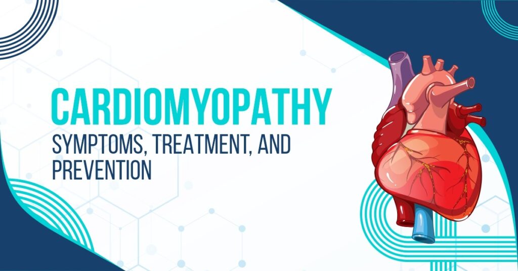
An enlarged heart muscle causes a condition called cardiomyopathy. The enlarged heart muscle stretches, which makes the muscle weak, thus preventing it from pumping blood as efficiently as it should.
You may develop heart failure if your heart muscle weakens too much (a serious condition that requires special treatment). Cardiomyopathy is relatively mild and most people can lead almost normal lives.
Heart attacks and cardiomyopathy are both conditions that damage a part of your heart muscle. Heart attacks may also cause you to need a heart transplant. People with severe heart failure may also need a heart transplant.
Types of Cardiomyopathy
Five types of cardiomyopathy are listed below:
- Dilated cardiomyopathy: A person with cardiomyopathy has an enlarged (dilated) left ventricle, thus limiting its ability to pump blood effectively.
This type of heart disease can affect anyone of any age, though it is more likely to affect men over the age of 50. It is most commonly caused by coronary artery disease or a heart attack, but it can also be caused by inherited defects.
- Hypertrophic cardiomyopathy: It is the type of cardiomyopathy in which an abnormal thickening of your heart muscle, which makes it more difficult for your heart to pump. It mainly occurs in the main pumping chamber of your heart (left ventricle).
Some genetic mutations have been linked to the development of hypertrophic cardiomyopathy. This condition can occur at any age but is typically more severe in childhood. People with this condition generally have a family history of it.
- Restrictive cardiomyopathy: It is the least common type of cardiomyopathy, characterized by stiffening and decreased flexibility of the heart muscle so it cannot expand and fill up with blood between heartbeats. Although it can affect people of any age, it most commonly affects older adults.
Occasionally, restrictive cardiomyopathy has no known cause (idiopathic), but it can also be caused by another disease elsewhere in the body that affects the heart, such as Amyloidosis.
- Arrhythmogenic right ventricular dysplasia: It is a rare type of cardiomyopathy caused by genetic mutations, causing scar tissue to replace the muscles in the lower right heart chamber (right ventricle).
- Unclassified cardiomyopathy: Other remaining types of cardiomyopathy can fall into this category.
Who is at risk for cardiomyopathy?
Risk factors for cardiomyopathy include certain diseases or conditions. Some of the most common are:
- Family History: Cardiomyopathy is more likely to occur in individuals with a history of it in their families.
- Hypertension: A person with long-standing hypertension who is not adequately controlled has a high risk of developing Cardiomyopathy.
- Thyroid Disorders: Cardiomyopathy is also common in people with thyroid problems.
- Diabetes Mellitus: Cardiomyopathy is also more common in diabetics with longstanding diseases.
- Obesity: Overweight individuals are at a greater risk of developing cardiomyopathy because their heart is put under more pressure.
- Alcoholism: Cardiomyopathy is also a risk factor for chronic alcoholics.
- Polysubstance Use: The risk of Cardiomyopathy increases in those who abuse drugs like amphetamines or cocaine as a recreational activity.
- Other Cardiac Condition: Cardiomyopathy can also affect individuals with preexisting cardiac conditions.
Symptoms of Cardiomyopathy
Common cardiomyopathy symptoms include:
- Unexplained fatigue
- Body weakness
- Sometimes feeling of fainting
- Regular chest pain
- Breathing problems
- Palpitations
- Fluid and water retention
- Light-headedness or headache
- Bloating
- Cough
- Dizziness
- Poor appetite
- Sudden death
- Swollen Extremities
How does cardiomyopathy affect children and teenagers?
There is no gender or race discrimination in pediatric cardiomyopathy for children or teenagers. Children and teenagers are more likely to develop this disorder when they are infants.
You can Read Also: BLADDER INFECTION SYMPTOMS, CAUSES & TREATMENT
In about 75% of cases, healthcare providers don’t know what causes cardiomyopathy in children. However, children may inherit cardiomyopathy or they may acquire it through a viral infection.
Early detection and treatment can improve a child’s outcome if cardiomyopathy is detected and treated during a sudden cardiac arrest, as some children may not exhibit symptoms.
The majority of children and teenagers with cardiomyopathy require routine medical attention from a cardiologist (a specialist in the heart); they receive daily medication. Depending on the cause, type, and stage of cardiomyopathy, there may not be many lifestyle restrictions.
Diagnose cardiomyopathy
In the diagnosis of the disease cardiomyopathy, the doctor will consider your medical history, family history to examine the exact cause of cardiomyopathy, physical examination, and diagnostic test results are used to diagnose cardiomyopathy. A cardiologist or pediatric cardiologist is often responsible for diagnosis and treatment. These doctors who specialize in heart disease are known as cardiologists or pediatric cardiologists.
- Medical and family histories: If any member of your family has been diagnosed with cardiomyopathy, heart failure, or cardiac arrest, your doctor will ask about your medical history and the signs and symptoms you are experiencing for further process of treatment.
- Physical exam: A stethoscope is used by your doctor to listen to the sounds of your heart and lungs to hear if they are similar to those that might indicate cardiomyopathy. Certain sounds may even indicate a specific type of the disease. A loud, rhythmic heart murmur can be a sign of obstructive hypertrophic cardiomyopathy. If a doctor hears a “crackling sound” in your lungs, this could symbolize heart failure. Your doctor can also use certain physical signs to detect cardiomyopathy. Ankle, foot, leg, abdomen, or neck swelling indicates fluid accumulation, a sign of heart failure.
- Diagnostic tests: To diagnose cardiomyopathy, your doctor might recommend any of these tests.
- Blood tests: By using a needle, blood is drawn from a vein in the arm to test.
- Chest X-ray: Images of your chest may reveal an enlarged heart or swelling of your lungs. An X-ray may also reveal fluid within your lungs.
- Electrocardiogram (EKG or ECG): The electrocardiogram records the heart’s electrical activity, showing how fast it beats and if its rhythm is steady or irregular. EKGs can help detect cardiomyopathy and other conditions such as heart attacks and arrhythmias (irregular heartbeats). Depending on the type of heart problem you have, your doctor may have you wear a portable EKG monitor.
- Holter and event monitor: In both cases, this is a portable device that monitors the heart’s electrical activity during a specific time. A Holter monitor provides long-term electrocardiogram data for 24 or 48 hours. An event monitor is limited to certain times only.
- Echocardiogram (Echo): Echocardiograms (echos) use sound waves to create a moving picture of your heart. They provide important information about how well your heart is working and its size and structure. Transesophageal echo (or TEE), which provides views of the back of the heart, is one type of echocardiography that is used during a stress test. Other types of echocardiography include those used during stress tests.
- Stress Test: In a stress test, your heart is systematically made to work hard (and beat fast) throughout various tests such as nuclear heart scanning, echocardiograms, and positron emission tomography (PET). During your appointment, you will walk on an inclined treadmill or receive medicine to simulate exertion if you are unable to exercise.
- Genetic Testing: Since Cardiomyopathy may also be genetically linked in some cases, the physician may suggest a genetic test if the patient has a family history of Cardiomyopathy.
Treatment for cardiomyopathy?
Treatment for cardiomyopathy involves controlling symptoms, stopping the progression of the disorder, and preventing sudden cardiac death. It can vary depending on the severity of symptoms and the type of cardiomyopathy.
Treatment of Cardiomyopathy includes:
Lifestyle changes:
The use of healthier lifestyle habits may also help slow the progression of cardiomyopathy. Lifestyle changes may help reduce the severity of conditions that may have caused the condition.
You can Read Also: BAD BREATH: CAUSES, TREATMENTS, AND PREVENTION
Following a healthful diet may also help modify lifestyle, as it limits the intake of saturated fats, trans fats, sugar, and salt.
Medications:
Some types of medicines may be prescribed to patients with cardiomyopathy, including:
- Beta-blockers: A beta-blocker slows the heart rate, resulting in less strain on the heart.
- Diuretics: Insufficient heart pumping can lead to excess fluid buildup in the body, which is removed by diuretics.
- Blood thinners: Blood-thinning medications lower your chance of developing blood clots.
- Blood pressure drugs: Heart disease patients can use angiotensin-converting enzyme (ACE) inhibitors, angiotensin receptor blockers, and angiotensin receptor-neprilysin inhibitors to reduce blood pressure and block the stress receptors.
- Antiarrhythmics: An antiarrhythmic is a medication that prevents abnormal heart rhythms.
Implanted devices:
Different types of implantable devices may also be used in treatment, depending on the symptoms. Implantable devices may include:
- Pacemaker: In such a case, a pacemaker can help the heartbeat at normal speed by delivering electrical impulses beneath the skin in the chest near the implantation site.
- Implantable cardioverter-defibrillator: As part of the device, on detection of an abnormal, potentially unstable heartbeat, the electric shock is delivered. This restores the heart rhythm to normal.
- Left ventricle assist device (LVAD): In this process, While a person receives a heart transplant, an LVAD is helpful while the heart is weakened by cardiomyopathy.
- Cardiac resynchronization device: This implanted device enhances heart function by improving coordination between left and right ventricles.
Surgery:
Surgery may be recommended if symptoms are severe. Possible surgery procedures for cardiomyopathy include:
- Septal myectomy: By removing the thickened tissue in the left ventricle, this surgery improves blood flow out of the heart. It is done for patients with hypertrophic cardiomyopathy with obstructions in blood flow.
- Heart transplant: Heart transplant might be an option for people who have certain types of cardiomyopathy with advanced heart failure. However, heart transplantation is a complex procedure, not everyone qualifies for one.
How can cardiomyopathy be prevented?
Cardiomyopathy can’t be prevented, but you can take steps to lower your risk for conditions responsible for creating (or complicating) cardiomyopathies, such as coronary heart disease, high blood pressure, and heart attacks.
An underlying health problem can precipitate cardiomyopathy. Treating it early can help prevent the complications associated with cardiomyopathy. As an example, you can control high blood pressure, high cholesterol, and diabetes by preventing the underlying causes:
- Regular checkups with your doctor are mandatory and all should do for their health.
- Follow your doctor’s advice about lifestyle changes and diet plans.
- Take all of your medications on time as prescribed by your doctor.
Cardiomyopathy can also cause other complications, just as some underlying conditions might cause cardiomyopathy.
Heart diseases, such as cardiomyopathy, can increase the risk for sudden cardiac arrest (SCA), which can be mitigated by implanted cardioverter defibrillators (ICDs).

