Introduction
Sialendoscopy is a minimally invasive procedure indicated for the diagnosis and treatment of salivary gland disorders which are commonly caused by obstructions like salivary stones, strictures, or infections. These conditions contribute to swellings, pain, and difficulties in saliva flow which commonly lead to debilitations of oral health and comfort.
In this article, we will discuss the different kinds of causes through which salivary gland obstruction occurs, its symptoms which reflect a medical problem, types of preventive measures available to avoid further glandular difficulties, and modes of treatment- one such an example being the sialendoscopy process, which seems to have impressive results. The factors listed out help in providing early diagnosis, and subsequently with management, make them suitable for better long-term oral health maintenance.
What is Sialendoscopy?
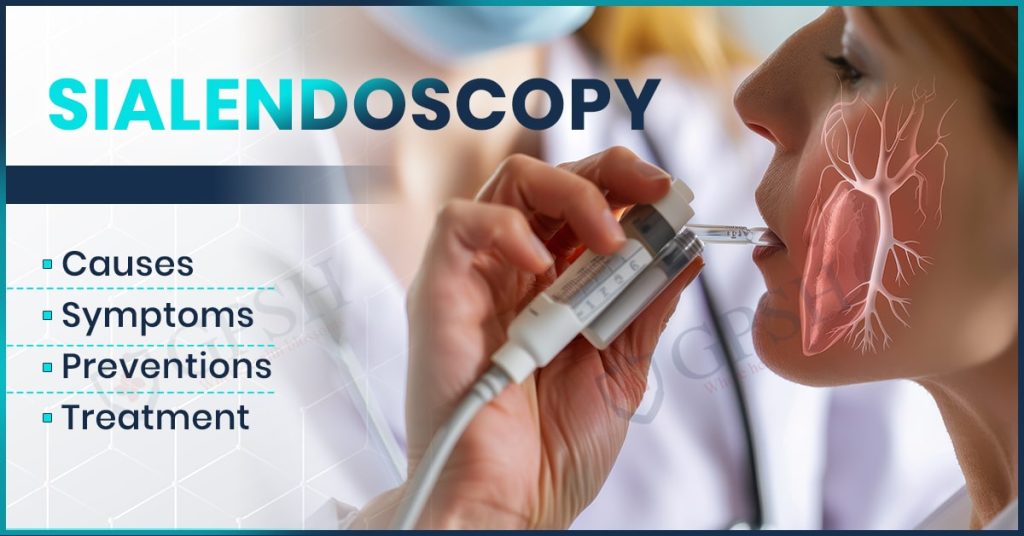
Sialendoscopy is a minimally invasive diagnostic and therapeutic procedure for examination and treatment of salivary duct disorders, including sialolithiasis, ductal strictures, and chronic infections. It involves the insertion of a miniature endoscope (0.8–1.6 mm in diameter) into the salivary ducts.
It allows for direct visualization and intervention without external incisions, maintaining gland function while enabling stone removal, ductal dilation, and lavage therapy. Biologically, sialendoscopy promotes the salivary gland by reinstating normal salivation flow and decreasing inflammation as well as the recurrence of infections.
You can read also:- Endocarditis: Causes, Symptoms, Risk Factors, and Treatment
Causes of salivary gland disorders
Here are the very few causes of Salivary Gland Disorders Leading to Sialendoscopy.
- Sialolithiasis (Salivary Stones): Calcium-based deposits obstruct salivary ducts, thus causing swelling and pain.
- Ductal Strictures: These are narrow bands of salivary ducts caused by scar tissue, inflammation, or recurrent infections.
- Chronic Sialadenitis: Recurrent infections from bacteria and viruses cause constant gland swelling and dysfunction.
- Autoimmune Disorders: Such conditions like Sjӧgren’s syndrome lead to decreased saliva secretion and duct blockages.
- Mucous Plugs: The plugs of thickened saliva or mucus block ducts, giving a sense of pain and inflammation.
- Trauma or Surgery: Ducts become scarred and obstructed by previous surgical interventions, injury, or radiotherapy.
- Dehydration and Impaired Saliva Flow: Lack of saliva raises the risk for stone formation as well as for infection.
- Drugs: Inhibitors to saliva production: antihistamines, diuretics, anticholinergics.
- Tumors or Cysts. Benign and malignant tumors might compress the ducts of their glands.
Symptoms of Salivary Gland Disorders
Here are some of the symptoms of Salivary Gland Disorder.
- Swelling of the Salivary Glands – Repeated or persistent swelling, often near the jaw, cheeks, or under the tongue.
- Pain or Tenderness – Pain, particularly when eating or drinking, as a result of duct obstruction.
- Dry Mouth (Xerostomia) – Decreased saliva production, causing difficulty in chewing, swallowing, or speaking.
- Obstruction of Saliva Flow – Feeling of blocked or decreased saliva production.
- Recurrent Infections – Infection of the ducts repeatedly by bacteria or viruses, giving rise to redness, warmth, and discharge of pus.
- Bad Taste or Foul Smelling Discharge – Pus or bacterial collections causing an objectionable taste or odor in the mouth.
- Hard Lumps or Stones in the Gland Area – Palpable stones present within the salivary ducts.
- Fever and General Malaise – Systemic signs of infection can occur, especially fever and generalized malaise.
- facial or jaw pain. It may radiate to other surrounding areas and may worsen with meals.
How is Sialendoscopy Performed?
Steps of Performing Sialendoscopy
-
Preparation of the Patient
- Local or general anesthesia is provided depending on the severity of the procedure.
- The salivary duct opening is softly dilated for scope placement.
-
Placement of Sialendoscope
- A micro-endoscope with a diameter of 0.8–1.6 mm is inserted slowly into the salivary duct for viewing purposes.
-
Duct Examination
- Camera and fiber-optic light give a magnified view of the interior of the duct. Any blockage present, such as stones, strictures, or infections, is detected.
-
Procedure for Treatment
- Salivary Stone Removal – The stones are removed using micro forceps, baskets, or laser fragmentation.Ductal Dilation – Tight strictures or narrowing is dilated by small balloon catheters.
- Lavage Therapy – Saline or anti-inflammatory solutions flush out debris and infection in the duct.
-
Final Inspection and Scope Removal
- The duct is checked for clearance and the scope is removed with care.
-
Post-Procedure Care
- Patients are monitored for swelling or discomfort.
- Hydration and gland massage are promoted to stimulate salivation.
- In case of necessary, antibiotics or pain relievers may be given.
You can read also:- Neck Pain in Children: Causes, Symptoms, and Treatment
Treatment Options for Salivary Gland Disorders
Disorders of the Salivary glands are caused due to infections, blockages, autoimmune diseases, or tumors. This condition depends upon the cause as well as on the severity; hence, some of the chief treatment options follow:
- Medications & Conservative management
- Antibiotics: When there is an infection caused by bacterial pathogens (for example sialadenitis).
- Anti-inflammatory & Analgesics- NSAIDs include ibuprofen which helps minimize pain and edema.
- Liquids and Sialogogues- More intake of fluid and sucking some sour candies sialogogues help stimulate salivary gland flow.
- Massage & Warm Compress: Facilitate dislodgement and allow gland drainage.
- Treatments of Obstruction
- Manual massage and hydration: Stones can be expectorated through the mouth
- Sialendoscopy: Minimally invasive removal of salivary stones
- Surgical removal: For large and embedded stones.
- Treatment of Autoimmune Conditions like Sjögren’s Syndrome
- Artificial saliva & eye drops: They facilitate moisturization.
- Medications: Pilocarpine or cevimeline increases the amount of saliva.
- Immunosuppressants: Steroids or biologics in severe cases
- Treatment of Tumors Benign or Malignant
- Surgical removal: First-line treatment for a tumor (such as pleomorphic adenoma)
- Radiation therapy: Malignant tumors, or if surgical intervention is not possible
- Chemotherapy: Not very common; it is utilized in aggressive and metastatic conditions
- Treatment of Viral Infections such as Mumps
- Supportive care: Rest, hydration, and pain control by NSAIDs or acetaminophen
- Isolation & prevention: Mumps vaccination, MM End.
Preventing Salivary Gland Issues
Here are some precautions you need to take so you do not get Salivary Gland Issues.
- Keep Your Mouth Hygienic: Brushing at least twice daily, flossing, and using an antibacterial mouthwash help prevent bacteria in the mouth. Poor oral hygiene can create a buildup of bacteria, which can travel down to the salivary glands and cause infection like sialadenitis which is inflammation of the salivary gland.
- Stay Hydrated: Drinking enough water throughout the day keeps the salivary glands active and prevents dehydration. A dry mouth increases the risk of bacterial infections and salivary gland blockages, which may lead to conditions such as xerostomia or dry mouth syndrome. Limit your intake of caffeine and alcohol, as these are dehydrating agents that decrease saliva production.
- Stimulate Saliva Flow: Chew sugar-free gum or eat foods high in fiber (such as apples, carrots, and celery) to naturally stimulate saliva. Saliva serves to wash out bacteria and food particles and also prevents the development of salivary stones.
- Avoid Smoking and excessive Alcohol. Smoking and alcohol irritate the salivary glands, increasing the chance of infection and blockage. These habits may also contribute to decreased saliva production, which can cause dry mouth and bacteria growth.
- Prevent Salivary Stones: Salivary stones, also known as sialolithiasis, are formed when saliva becomes concentrated, resulting in crystallized deposits in the glands. A balanced diet rich in fluids and low in excessive calcium intake helps prevent stone formation. Gently massaging the salivary glands can promote saliva flow and reduce blockages.
- Manage Underlying Conditions: Several medical conditions lead to decreased production of saliva by the salivary glands; these include diabetes, Sjögren’s syndrome, and autoimmune diseases. Management of the underlying conditions either through medication or lifestyle adjustments safeguards the salivary glands against dysfunction.
- Protect Against Viral Infections: Viral infections, including mumps (which are caused by the mumps virus), influenza, and cytomegalovirus, directly affect the salivary glands. Getting vaccinated with the MMR vaccine (measles, mumps, and rubella) and maintaining good hygiene (frequent handwashing and avoiding contact with infected individuals) helps prevent these infections.
Conclusion
Salivary gland disorders impair oral and general health, often resulting from infections, blockage, autoimmune conditions, or sometimes due to tumors. Of the most advanced and minimally invasive procedures to treat salivary gland disease, especially salivary duct stones, is sialendoscopy.
This procedure lets a doctor come to your rescue with precise diagnosis and treatment that brings less discomfort and quicker recovery than surgery. Salivary gland disorders and their treatments are undertaken in the departments of Otolaryngology (ENT) and Oral & Maxillofacial Surgery, where specialized care is provided. Shekhawati Hospital has a reputation for diagnosing and treating salivary gland conditions with advanced procedures such as sialendoscopy to ensure effective and patient-friendly treatment.